Deep Vein Thrombosis (DVT) Ultrasound Scan

Deep Vein Thrombosis (DVT) Ultrasound Scan

Deep vein thrombosis (DVT) is a blood clot that develops within a deep vein of the body, usually in the leg and ultrasound scans are commonly used to diagnose or exclude the existence of DVT. DVT usually occurs in a deep leg vein, a larger vein that runs through the muscles of the calf and the thigh.
It can cause pain and swelling in the leg and may lead to complications such as pulmonary embolism. This is a serious condition that occurs when a piece of blood clot breaks off into the bloodstream and blocks one of the blood vessels in the lungs (see below).

The DVT ultrasound scan is the first-line imaging technique to check the veins for any signs of a clot.

WHAT OUR PATIENTS SAY

What is the cost of the DTV scan?

The cost of this ultrasound scan is only £164 (both legs £235) .

Book Your  DVT Ultrasound Appointment Today

Symptoms of DVT

In some cases, there may be no symptoms of DVT.

If symptoms do occur they can include:

  • pain,
  • swelling and tenderness in one of your legs (usually your calf)
  • a heavy ache in the affected area
  • warm skin in the area of the clot, particularly at the back of your leg below the knee

DVT usually (although not always) affects one leg. The pain may be worse when you bend your foot upward towards your knee.


What causes DVT?

Each year, DVT affects around one person in every 1,000 in the UK. Anyone can develop DVT, but it becomes more common over the age of 40. As well as age, there are also a number of other risk factors, including:

  • having a history of DVT or pulmonary embolism
  • a family history of blood clots being inactive for long periods – such as after an operation or during a long journey
  • blood vessel damage – a damaged blood vessel wall can result in the formation of a blood clot
  • having certain conditions or treatments that cause your blood to clot more easily than normal – such as cancer (including chemotherapy and radiotherapy treatment),
  • heart and lung disease,
  • thrombophilia and Hughes syndrome
  • being pregnant – your blood also clots more easily during pregnancy
  • being overweight or obese
  • The combined contraceptive pill and hormone replacement therapy (HRT) both contain the female hormone oestrogen, which causes the blood to clot more easily. If you're taking either of these, your risk of developing DVT is slightly increased.


What is the purpose of the DVT ultrasound scan?

The DVT ultrasound scan includes evaluation of:

  • Femoral vein
  • Saphenofemoral junction
  • Popliteal vein
  • Deep calf veins (if visualized)


Reasons for having a DVT ultrasound scan:

  • Pain,
  • swelling and tenderness in one of your legs (commonly in the calf region)
  • A heavy ache in the affected area.
  • Warm skin in the area of the clot.
  • Redness of your skin, particularly at the back of your leg below the knee.


Preparation

No preparation for this private DVT ultrasound scan is necessary.


Price: £164.00 (both legs £235)

Private  Ultrasound 

Do you need an ultrasound and you do not want to wait 6+ weeks for a hospital one?

Book Your Scan Now

What should I expect during the DVT ultrasound scan?

Upon your arrival, our sonographer will explain the ultrasound scan procedure. You will need to expose your legs and a small amount of gel will be placed on your skin. The sonographer will follow the length of your leg veins and from time to time you will feel a small amount of pressure. 


About Vascular Ultrasound

Vascular ultrasound is carried out in order to monitor blood flow to organs and tissues throughout the body, locate and identify blockages and abnormalities like blood clots, plaque or emboli.

Vascular ultrasound can also help to identify areas of an abnormal widening of blood vessels (an aneurysm) that, if left untreated, can lead to serious consequences.
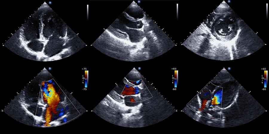
Doppler ultrasound study may be part of a vascular ultrasound examination. Doppler is a special ultrasound technique that evaluates the speed and volume of blood as it flows through a blood vessel, including the body's major arteries and veins in the abdomen, arms, legs, and neck. It can help to diagnose blockages to blood flow (such as clots) narrowing of vessels (possibly caused by plaque) and tumours and congenital malformation.
As the images are captured in real-time, they can help the sonographer monitor the blood flow to organs and tissues throughout the body.


Price: £164.00 (both legs £235)

Book Your  Appointment Now

Book your appoinment online.
BOOK AN APPOINTMENT

DVT Scan Q&A


What are ultrasound scans?

Ultrasound imaging is a medical diagnostic technique where sound waves are being used to image various parts of the body.

Other terms for ultrasound imaging that are being used are sonograms, US and sonography.
Ultrasound is widely used these days as it is painless and safe to adults, children and foetuses. There are no side effects such as the ones associated with ionising radiation produced by CT and plain x-rays.
During the ultrasound scan, the sonographer rests a small probe over the skin. This probe produces sound waves i.e. pulsations that travel through your skin and inside your body. Part of the sound waves are being reflected back to the transducer and the computer analyses the returning echoes and produces the image on the screen. Medical ultrasonography uses the same principle as the sonar that the navy uses to detect submarines.
Ultrasound is being used to image mostly solid soft organs such as liver, kidneys, uterus and ovaries, muscles and blood vessels and babies in the womb. It has, however, limited value in organs such lungs, bone, stomach and bowel/colon.
Ultrasound images are black and white but colour Doppler is being used to evaluate organ blood flow and blood vessels and this is what the red and blue colours on the screen are.


Looking for a DVT ultrasound scan in London?

Our private scan clinic specialises in diagnostic private ultrasound to provide you with quick diagnosis and the much-needed reassurance about your health, within a convenient location. The cost of a private ultrasound scan shouldn’t be prohibited when it comes to someone’s wellbeing. This is why our prices are competitive and affordable with no compromise to the level of care. The price you see is the price you pay with no hidden extras. We have hundreds of happy clients including GP’s, consultants, radiologists and midwives to name a few. We can conveniently book an ultrasound scan appointment via our online booking system or on the phone.


What are the benefits and risks of the DVT ultrasound examination?

Benefits

  • Ultrasound scans are non-invasive.
  • An ultrasound exam should not be painful.
  • Ultrasound is widely available, easy-to-use and less expensive than other imaging modalities.
  • Ultrasound imaging is extremely safe and does not use any harmful ionizing radiation and this is why is the primary imaging modality in pregnancy.
  • Ultrasound scanning gives a clear picture of soft tissues and blood vessels that do not show up well on x-rays.
  • Venous ultrasound helps to detect blood clots in the veins of the legs before they become dislodged and pass to the lungs. It can also show the movement of blood within blood vessels.

Risks

  • There are no known harmful effects on humans related to ultrasound scans.
 

Who interprets the results of the DVT scan and how do I get them?

Our Sonographer, a Health Care Professional specifically trained to perform and understand the ultrasound scan images, will do your ultrasound and provide you with an ultrasound report that you can take it your doctor.  Our sonographers will also discuss the results with you during and after your examination.

Our sonographers are advanced consultant practitioners with years of ultrasound scanning experience. They are fully qualified, registered with the HCPC, BMUS and SoR and work full/part time for the NHS and private clinics. On an average month, they perform more than 500 examinations and undertake regular CPD and other training activities to keep up with advances in ultrasound scanning technology. You can, therefore, you can rest assured that you are in good hands.


What causes Deep Vein Thrombosis?

According to NHS Choices:

Each year, DVT affects around one person in every 1,000 in the UK.
Anyone can develop DVT, but it becomes more common over the age of 40. As well as age, there are also a number of other risk factors, including:

  • having a history of DVT or pulmonary embolism
  • having a family history of blood clots
  • being inactive for long periods – such as after an operation or during a long journey
  • blood vessel damage – a damaged blood vessel wall can result in the formation of a blood clot
  • having certain conditions or treatments that cause your blood to clot more easily than normal – such as cancer (including chemotherapy and radiotherapy treatment), heart and lung disease, thrombophilia and Hughes syndrome
  • being pregnant – your blood also clots more easily during pregnancy
  • being overweight or obese

The combined contraceptive pill and hormone replacement therapy (HRT) both contain the female hormone oestrogen, which causes the blood to clot more easily. If you're taking either of these, your risk of developing DVT is slightly increased.

What are the limitations of DVT Ultrasound scan Imaging?

It can be hard to fully visualise the internal anatomy of large patients as the sound has further to travel and therefore the returning echoes are weaker

Veins lying deep beneath the skin, especially small veins in the calf, may be hard to see.

Venous ultrasonography is accurate for detecting blood clots in the veins of the upper leg (duplex ultrasound identifies 95% of the clots) but is less accurate in the calf veins, as the veins become very small. DVT ultrasound identifies up to 70% of calf vein DVT. Potentially dangerous venous clots are however typically found in the larger femoral veins and calf vein thrombi are less likely to break and travel to the lungs to cause pulmonary embolism


Why choose us for your private DVT ultrasound scan in London?


We are conveniently located in the heart of London, in Harley street just a few minutes’ walk from Oxford Circus station. 
We offer same-day appointments in a clean and caring environment.

We have years of experience in medical ultrasound. Experience gained working for flagship NHS trusts alongside leaders in the field of diagnostic medical imaging including general ultrasound, urology, musculoskeletal, gynaecology, pelvis, testicular and vascular examinations. You can, therefore, be assured that your health is in good hands.

We have thousands of happy clients and five-star reviews on independent websites.

Ultrasound London Clinic Address:
29 Weymouth Street, Marylebone, London, W1G 7DB
Phone: 020 3633 4902 Email: info@sonoworld.co.uk or sonoworlduk@gmail.com
calendar-fullcrossmenu Tap to Call