
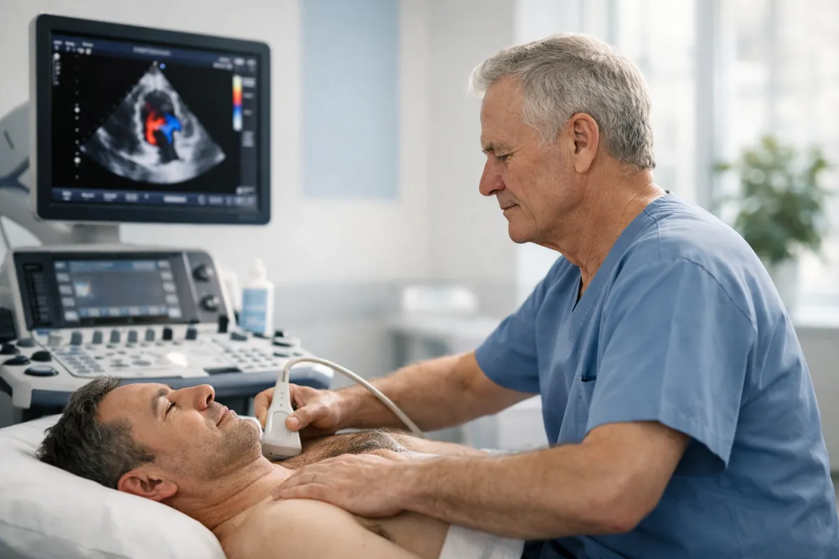
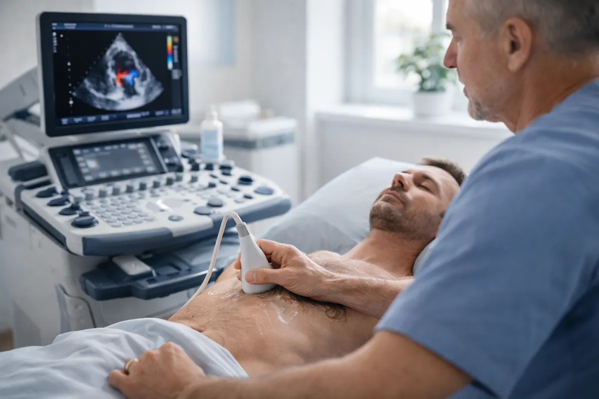
Quick definition: what is an ultrasound echo?
An ultrasound echo—also called an echocardiogram—is a medical ultrasound test that uses sound waves to create real-time images of the heart. It helps clinicians evaluate:
- Heart chambers (size and shape)
- Heart muscle function (how well it pumps)
- Valves (whether they open/close properly, narrowing or leakage)
- Blood flow through the heart and major vessels using Doppler
- Pericardium (fluid around the heart)
Types of echocardiogram: TTE vs TEE (and Doppler)
Most private “ultrasound echo” appointments are Transthoracic Echocardiograms (TTE)—a probe on the chest. Some hospital pathways use Transoesophageal Echocardiograms (TEE) for specific indications.
| Type | How it’s done | Typical use | Comfort |
|---|---|---|---|
| TTE (standard echo) | Probe on the chest with gel. Non-invasive. | First-line imaging for function + valves + fluid assessment. | Comfortable |
| TEE (specialised) | Probe passed into the oesophagus (sedation often used). | When very detailed valve/structure views are needed. | More invasive |
| Doppler / Colour Doppler | Mode used during TTE/TEE to measure blood flow direction & velocity. | Valve leakage/narrowing, pressure estimates (where clinically valid). | Part of scan |
How does cardiac ultrasound work?
Ultrasound uses high-frequency sound waves that reflect (“echo”) off tissues. The machine converts returning echoes into images. In the heart, this allows moving pictures of valves and heart muscle. Doppler ultrasound measures changes in frequency of echoes from moving blood—helping assess flow direction and speed.
Why operator skill matters
Echocardiography depends on finding the correct acoustic windows and optimising settings (gain, depth, Doppler angles). This is why training and structured reporting are important—especially for valve severity and functional measurements.
- Better windows = clearer valve and wall motion assessment
- Doppler alignment influences flow velocity accuracy
- Structured measurements improve clinical interpretability
Booking at Sonoworld: Echocardiogram appointments
What does an echo check in a structured exam?
A clinically useful echo is organised by entities and attributes—so it answers questions clinicians act on. Typical reporting domains include:
Chambers
Left/right ventricle and atria size, geometry, wall thickness.
Function
Global function (e.g., EF where measurable), wall motion patterns.
Valves
Stenosis/regurgitation screening; Doppler flow assessment.
Pericardium
Fluid around the heart (pericardial effusion) and effects.
Aorta
Aortic root/ascending aorta visualisation where windows allow.
Doppler
Flow direction/velocity; supports pressure estimates in context.
When is an ultrasound echo recommended?
Echocardiography is commonly used to investigate symptoms, assess a murmur, monitor known conditions, or establish a baseline. Some of the most frequent indications include:
- Breathlessness, reduced exercise tolerance, or unexplained fatigue
- Heart murmur or suspected valve disease
- Palpitations (often alongside ECG/Holter monitoring)
- Swollen ankles/legs with a cardiac query (may overlap with vascular causes)
- Follow-up for known diagnoses (e.g., valve disease, cardiomyopathy) as advised by a clinician
Echo vs ECG vs CT vs MRI: which test answers which question?
The most common booking confusion is expecting one test to do everything. Here’s a practical comparison based on clinical “jobs to be done.”
| Test | Best for | Limitations | Radiation? |
|---|---|---|---|
| Echocardiogram (Echo) | Structure + function, valves, pericardium, Doppler flow. | Does not directly image coronary artery blockages reliably. | No |
| ECG | Electrical rhythm, conduction, acute changes. | Not an imaging test; doesn’t show valve structure or pumping directly. | No |
| Cardiac CT | Coronary anatomy (selected pathways), detailed anatomy. | Radiation exposure; contrast considerations; not first-line for valves. | Yes |
| Cardiac MRI | Tissue characterisation, complex cardiomyopathy assessment. | Longer exam; availability; not always first-line for urgent questions. | No |
If you’re uncertain which test best matches your symptoms, call 020 3633 4902 for guidance.
Is an ultrasound echo safe? Risks and reassurance
A standard transthoracic echo (TTE) is considered very safe. It uses ultrasound (sound waves), not ionising radiation. Most people experience no discomfort beyond mild pressure from the probe and cool gel.
Radiation
No radiation. Safe for repeated use when clinically indicated.
Discomfort
Usually minimal: gentle pressure and cool gel.
TEE caveat
TEE is more invasive and may involve sedation; it is used for specific clinical needs.
Understanding results: common terms you may see
Echo reports use clinical language so your GP or cardiologist can act on the findings. Common terms include:
- Ejection fraction (EF): a measure of left ventricular pumping performance (interpreted in context).
- Regurgitation: valve leakage (mild/moderate/severe grading depends on multiple parameters).
- Stenosis: valve narrowing (assessed with Doppler velocities and calculated areas where relevant).
- LVH: left ventricular hypertrophy (thickened heart muscle, often related to blood pressure and other causes).
- Pericardial effusion: fluid around the heart (size and haemodynamic impact matter).
What happens at a private echocardiogram appointment?
Most private echoes follow a simple pathway: book → attend → scan + focused history → receive report with next-step guidance. Sonoworld accepts self-referrals and medical insurance.
Practical checklist
- Wear a 2-piece outfit for convenience.
- Bring medications list and any relevant prior results (if available).
- If using insurance: confirm coverage and bring authorisation details if required.
Also see: Carotid ultrasound • DVT ultrasound • AAA scan


