Specialist Doppler ultrasound assessment of the carotid arteries for stenosis, plaque, and stroke risk. CQC-registered Harley Street clinic, same-day appointments, no GP referral required.
Instant verbal results. Written report within 24 hours.
The carotid arteries are the two main blood vessels in the neck that supply blood to the brain. Atherosclerosis — the build-up of fatty plaque — within the carotid arteries is a leading cause of ischaemic stroke and transient ischaemic attack (TIA). A carotid Doppler scan uses high-resolution ultrasound and Doppler imaging to assess the carotid arteries for stenosis, plaque, and arterial wall thickening.
Carotid ultrasound is the first-line investigation recommended for stroke risk assessment and is used to guide decisions about medical management and surgical intervention (carotid endarterectomy). It is also used as a preventive screening tool to assess subclinical atherosclerosis in high-risk individuals.
Measurement of the carotid intima-media thickness — a validated marker of subclinical atherosclerosis and an independent predictor of future cardiovascular events. Increased IMT is associated with elevated risk of myocardial infarction and stroke.
Identification and characterisation of atherosclerotic plaque at the carotid bifurcation and internal carotid artery — the most common site of clinically significant stenosis. Plaque morphology (echogenicity, surface irregularity) is assessed to stratify embolic risk.
Colour Doppler and spectral waveform analysis to measure peak systolic velocity (PSV) and end-diastolic velocity (EDV) at the internal carotid artery. These measurements are used to grade the degree of stenosis according to established criteria (NASCET/ECST).
A carotid Doppler scan is appropriate for anyone with symptoms suggesting cerebrovascular disease, or for individuals who wish to assess their cardiovascular risk proactively. It is also included as part of our Cardiovascular Screening Package.
Our specialist sonographer takes a brief clinical history including your symptoms, cardiovascular risk factors, and any previous investigations. This ensures the examination is tailored to your clinical picture.
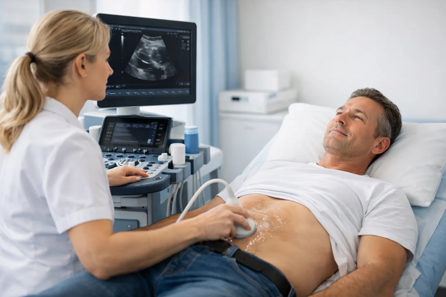
You lie on the examination couch with your head turned slightly to one side. Gel is applied to the neck and the sonographer uses a high-frequency probe to assess both carotid arteries systematically from the common carotid to the internal and external branches.
Your sonographer explains their findings immediately after the scan, including any plaque identified, IMT measurements, and the degree of stenosis if present. You can ask questions about what the findings mean for your management.
A comprehensive written report is emailed to you within 24 hours. The report includes IMT measurements, plaque characterisation, stenosis grading, and clinical recommendations. It can be shared directly with your GP, neurologist, or vascular surgeon.
Also included in our Cardiovascular Screening Package (£235 — echocardiogram + carotid + AAA). See also: Cardiovascular Scans Hub
No. You can self-refer directly online or by calling 020 3633 4902. If your GP has referred you following a TIA or carotid bruit, please bring the referral letter as it helps guide the examination.
A carotid bruit is an abnormal sound heard through a stethoscope over the carotid artery, caused by turbulent blood flow. It may indicate carotid artery stenosis. Not all bruits indicate significant stenosis, and not all significant stenoses produce a bruit — which is why ultrasound assessment is required for accurate diagnosis.
NICE guidelines recommend carotid endarterectomy (surgical removal of plaque) for symptomatic patients with 50–99% stenosis of the internal carotid artery, ideally within 14 days of a TIA or minor stroke. The decision is made by a vascular surgeon based on the full clinical picture, including the ultrasound findings.
Yes. Carotid ultrasound is the primary imaging tool for assessing stroke risk from carotid artery disease. Intima-media thickness measurement and plaque characterisation provide independent risk stratification beyond traditional cardiovascular risk factors. However, carotid ultrasound does not assess all causes of stroke — cardioembolic stroke (from the heart) requires an echocardiogram.
Carotid ultrasound is non-invasive, uses no radiation, and provides real-time dynamic assessment of blood flow. It is the first-line investigation for carotid artery disease. MRI and CT angiography provide additional anatomical detail and are used for surgical planning or when ultrasound findings are inconclusive.