
- Home
- Women
- Men
- MSK
- Cardiovascular
- Screening
- About
- Book a Scan
- Blog
A comprehensive vascular ultrasound-scan package assessing the carotid arteries, abdominal aorta, and bilateral leg arteries — the three most clinically significant sites for stroke and cardiovascular risk. One appointment, same-day written report.
Stroke is the fourth leading cause of death in the UK and the single largest cause of complex disability. Around 100,000 people have a stroke each year in England, and up to 80% of strokes are preventable when vascular risk factors are identified and managed early. The Sonoworld Vascular Stroke Screening package uses high-resolution colour Doppler ultrasound to examine the three arterial territories most directly linked to stroke and cardiovascular events.
Unlike a single-site vascular scan, this package combines three examinations into one 45-minute appointment. The carotid duplex assesses plaque burden and stenosis in the neck arteries supplying the brain. The abdominal aortic aneurysm (AAA) scan measures aortic diameter to detect aneurysmal dilatation before rupture. The bilateral leg arterial evaluation identifies peripheral arterial disease (PAD), which shares the same atherosclerotic pathology as carotid disease and is a strong independent predictor of future cardiovascular events.
Atherosclerosis — the build-up of fatty plaques in artery walls — is a systemic disease. Plaque in the carotid arteries frequently coexists with aortic dilatation and peripheral arterial disease. Examining all three territories in one appointment gives a complete picture of your systemic vascular health, rather than a partial view from a single-site scan.
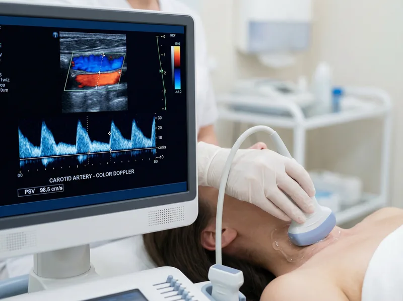
Each of the three examinations uses high-frequency B-mode imaging combined with colour Doppler and spectral waveform analysis to characterise both vessel anatomy and blood flow dynamics.
The carotid arteries are the primary blood supply to the brain. Atherosclerotic plaque at the carotid bifurcation — where the common carotid artery divides into the internal and external carotid — is the most common source of embolic stroke. The carotid duplex assesses:
An abdominal aortic aneurysm is a localised dilatation of the aorta exceeding 3 cm in diameter. AAAs are largely asymptomatic until rupture, which carries a mortality rate exceeding 80%. The NHS AAA screening programme invites men at age 65, but women and younger men with risk factors receive no routine NHS screening. This scan assesses:
Peripheral arterial disease (PAD) affects approximately 20% of people over 60 in the UK. Most cases are asymptomatic or present only with exertional leg pain (intermittent claudication). PAD is a powerful marker of systemic atherosclerosis — people with PAD have a two- to three-fold increased risk of myocardial infarction and stroke compared to those without. This evaluation assesses:
Vascular stroke screening is most valuable for people who carry one or more established cardiovascular risk factors but have no current symptoms. The screening identifies subclinical disease — arterial changes that are present and progressing but have not yet caused a TIA, stroke, or other vascular event.
Sudden facial drooping, arm weakness, or speech difficulty (FAST symptoms); sudden severe headache; sudden loss of vision in one eye; or sudden loss of balance. These are symptoms of an active stroke or TIA and require immediate emergency care — call 999, not a screening clinic.

The table below maps each scan area to the conditions it detects and the clinical action that follows a positive finding.
| Scan Area | Condition Detected | Clinical Significance | Action if Found |
|---|---|---|---|
| Carotid Duplex | Carotid stenosis ≥50% | Embolic stroke risk increases proportionally with stenosis severity; ≥70% stenosis may warrant endarterectomy | Urgent vascular surgery referral; antiplatelet therapy review |
| Carotid Duplex | Soft (echopenic) carotid plaque | Unstable plaque with higher embolic potential than calcified plaque, even at moderate stenosis | Intensified statin therapy; GP/cardiologist review |
| Carotid Duplex | Increased CIMT (>0.9 mm) | Subclinical atherosclerosis; independent predictor of future MI and stroke | Lifestyle modification; lipid-lowering therapy; GP review |
| AAA Scan | Aortic diameter 3.0–4.4 cm (small AAA) | Surveillance required; annual growth rate ~2–3 mm/year | Vascular surgery referral; 12-monthly surveillance |
| AAA Scan | Aortic diameter 4.5–5.4 cm (medium AAA) | Rupture risk ~1% per year; 3-monthly surveillance indicated | Urgent vascular surgery referral; 3-monthly surveillance |
| AAA Scan | Aortic diameter ≥5.5 cm (large AAA) | Rupture risk ~25% per year; surgical repair indicated | Same-day urgent vascular surgery referral |
| Leg Arterial | Peripheral arterial disease (PAD) | 2–3× increased risk of MI and stroke; limb ischaemia risk if untreated | GP referral; antiplatelet therapy; cardiovascular risk factor management |
| Leg Arterial | Popliteal aneurysm | Thrombosis and distal embolisation risk; limb-threatening if untreated | Vascular surgery referral |
The entire package is completed in a single 45-minute appointment by an HCPC-registered vascular sonographer. No GP referral is required.
The sonographer takes a brief cardiovascular history — symptoms, risk factors, current medications, and family history. This guides the clinical focus of the examination and ensures all relevant areas receive appropriate attention.
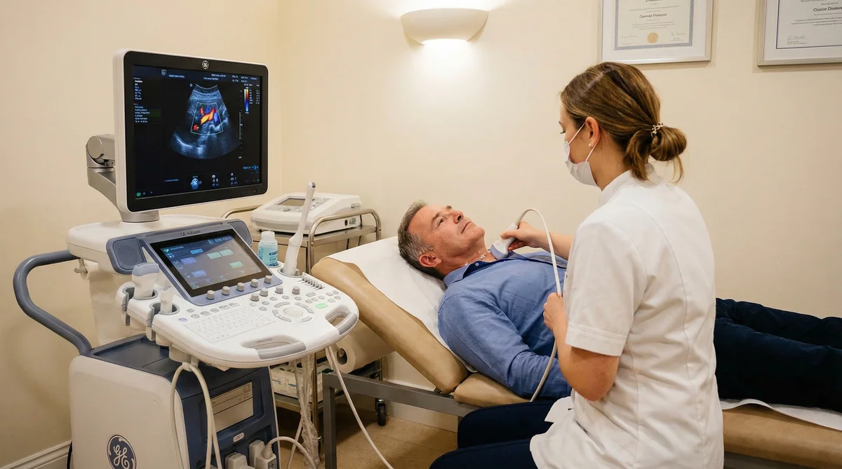
You lie on your back with your head turned to one side. The sonographer applies gel to the neck and uses a high-frequency linear probe to examine both carotid arteries and vertebral arteries. Colour Doppler and spectral waveform analysis are performed at each key site.
The probe moves to the abdomen. The aorta is measured at multiple levels from the diaphragm to the bifurcation. Colour Doppler assesses flow patterns. The iliac arteries are also evaluated. A 4-hour fast is required for this part of the examination to reduce bowel gas interference.
Both legs are examined from the groin to the ankle. The sonographer assesses the femoral, popliteal, and tibial arteries using B-mode imaging and Doppler waveform analysis. You will be asked to lie on your front for the popliteal assessment.
Findings are explained verbally at the end of the appointment. A comprehensive written report is sent to you — and to your GP or specialist if requested — the same day. The report includes measurements, images, and clinical recommendations for any abnormal findings.
The abdominal aorta assessment requires a 4-hour fast to minimise bowel gas. The carotid and leg arterial components require no special preparation.
Do not eat for at least 4 hours before your appointment. You may drink water freely — staying well hydrated actually improves image quality. Avoid fizzy drinks, which increase bowel gas.
Take all regular medications as normal, including antihypertensives, statins, and antiplatelet agents. Do not stop any medication before the scan.
Wear loose, comfortable clothing. A two-piece outfit is ideal — the sonographer will need access to your neck, abdomen, and both legs. Avoid tight trousers or leggings that are difficult to roll up.
One all-inclusive price for all three vascular examinations, verbal feedback, and a same-day written report.
Standalone carotid artery assessment — plaque, stenosis, CIMT, and vertebral artery flow.
Standalone AAA ultrasound-scan measuring aortic diameter and assessing aneurysm risk.
Comprehensive 6-area package including thyroid, carotid, abdomen, urinary tract, aorta, and testes.
No GP referral is required. You can book directly online or by telephone. If you have a GP referral letter, bring it to the appointment — it provides useful clinical context. The report is sent to your GP automatically if you provide their details.
Ultrasound is entirely non-invasive and painless. The probe is pressed gently against the skin with a small amount of warm gel. The only mild discomfort some patients experience is during the popliteal (behind-knee) assessment, which requires lying face-down for a few minutes.
The full package takes approximately 45 minutes. Allow a little extra time for the clinical history at the start and for verbal feedback at the end. You will not need to stay after the appointment — the written report is sent electronically the same day.
The sonographer will explain any findings verbally during the appointment. The written report includes clinical recommendations — for example, GP review, specialist referral, or repeat surveillance at a specified interval. For findings that require urgent action (such as a large aortic aneurysm), the sonographer will advise you to seek same-day medical review and will contact your GP directly if you consent.
Yes. Although the NHS AAA screening programme targets men aged 65, women with cardiovascular risk factors — particularly hypertension, smoking history, or a family history of AAA — also benefit from vascular screening. Carotid disease and peripheral arterial disease affect both sexes. The package is available to all adults.
Sonoworld is recognised by most major private health insurers. Contact your insurer before booking to confirm coverage and obtain an authorisation code. Bring the code to your appointment. If your insurer does not cover screening, self-pay is available at the package price of £699.
If the initial screening is entirely normal and your cardiovascular risk factors are well controlled, a repeat screen every 3–5 years is reasonable. If the scan identifies mild disease — such as early plaque or a small AAA — the report will specify the recommended surveillance interval. Your GP or vascular specialist can advise on the appropriate frequency based on your overall cardiovascular risk profile.
Address:
29 Weymouth Street, Marylebone
London W1G 7DB
Telephone:
020 3633 4902
Email:
info@sonoworld.co.uk
Opening Hours:
Monday – Friday: 8:00 am – 7:00 pm
Saturday: 9:00 am – 5:00 pm
Sunday: Closed


