
- Home
- Women
- Men
- MSK
- Cardiovascular
- Screening
- About
- Book a Scan
- Blog
Choose your appointment time below. No GP referral is required. Your scan takes approximately 20 minutes and your written report is ready within 24 hours.
The abdominal aorta lies deep in the abdomen, and bowel gas is the main obstacle to a clear image. You do not need to fast completely, but avoiding gas-producing foods for 24 hours before your scan significantly improves image quality.
Ultrasound is regularly being used to evaluate abdominal aortic aneurysms. Abdominal aneurysm is a swelling of the aorta, the main blood vessel that brings blood for your heart to the rest of the body. The aorta is around 2.5 in the abdominal area, it can, however, swell to more than 5 cm.
Aneurysms are more common in men of 65+ years of age than in women and younger men, and they can be fatal as large aneurysms can burst. According to NHS Choices 8 out of 10 people with aortic aneurysm or AAA die.
It is highly unlikely that small aneurysms will cause any symptoms but large aneurysm can cause the following symptoms:
Your sonographer will give you verbal feedback immediately. Your written report — including aortic measurements and clinical findings — will be sent to you within 24 hours.
If your aorta measures 5.5 cm or more, your sonographer will advise you to seek urgent vascular surgical review.
From confirmation to report — here is exactly what to expect at every stage of your AAA ultrasound-scan appointment.
You will receive an email confirmation immediately after booking, including your appointment time, address, and preparation instructions. A reminder is sent 24 hours before your appointment.
The clinic is at 29 Weymouth Street, Marylebone, London W1G 8GR — a 5-minute walk from Regent's Park and Great Portland Street tube stations. Arrive 5 minutes early to complete a brief patient history form.
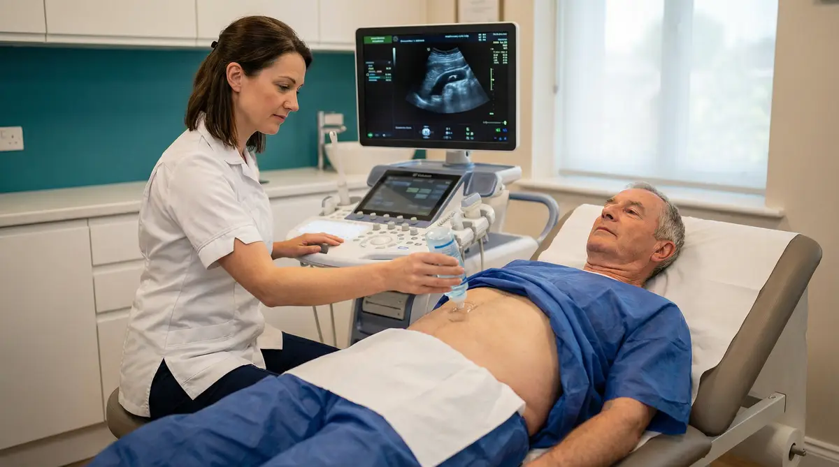
Your HCPC-registered sonographer will review your history, discuss your symptoms or risk factors, and confirm the scan protocol. You will be asked to lie on the examination couch and expose your abdomen.
Warm gel is applied to your abdomen. The sonographer uses a linear or curvilinear probe to image the aorta from the diaphragm to the bifurcation, measuring diameter in both transverse and longitudinal planes. Colour Doppler confirms flow patterns and identifies any mural thrombus or calcification.
Your sonographer will give you verbal feedback before you leave. Your written report — with aortic measurements, NICE NG156 classification (normal / small / medium / large), and clinical recommendations — is sent to you within 24 hours. If urgent findings are identified, you will be advised immediately.
29 Weymouth Street
Marylebone
London W1G 8GR
Phone: 020 3633 4902
Email: info@sonoworld.co.uk
Open in Google Maps| Monday – Friday | 8:00 am – 6:00 pm |
| Saturday | 9:00 am – 2:00 pm |
| Sunday | Closed |


