

Palpitations
Fast, fluttering, or irregular heartbeat
An ECG can capture rhythm abnormalities if they’re present during the test. If symptoms come and go, a Holter monitor can be a better match.
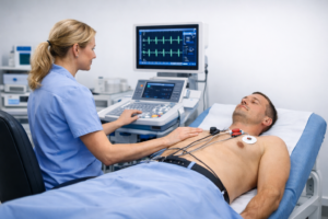
An ECG is a quick, non-invasive test that records the electrical activity of your heart. It helps us assess symptoms like palpitations, chest discomfort, dizziness, or an “irregular heartbeat” feeling — and decide whether you need reassurance, treatment, or the next test (like a Holter monitor or an echo).
If you have severe chest pain, breathlessness at rest, fainting, or new neurological symptoms, seek urgent medical help (A&E/999). For non-urgent concerns, an ECG is often the fastest first step.
An ECG (electrocardiogram) records your heart’s electrical signals through small skin electrodes. Think of it as a “rhythm snapshot” — it can show if the timing and pathways of the electrical impulses look normal during the recording.
If your main concern is “Do I need an echo?” start with our triage page: Symptoms that require an echo.

In real life, most people don’t search for “ECG” first. They search for the feeling: a flutter, a missed beat, a tight chest, or light-headedness. An ECG is usually the quickest way to turn that worry into a clinical plan.
An ECG can capture rhythm abnormalities if they’re present during the test. If symptoms come and go, a Holter monitor can be a better match.
An ECG is often part of an initial assessment. If your symptoms suggest a structural or pumping issue, we may recommend an echo as well.
An ECG can identify conduction patterns that may explain symptoms. If episodes are intermittent, longer monitoring is often more informative.
Use the triage page to decide what to book first — it’s designed around real patient journeys (what you feel → what that can mean → best next test).
If you’re anxious, the most helpful thing to know is this: the test is quick, and nothing “goes into” your body. We’re simply reading signals from the skin.
Here’s the simplest way to decide: ECG is best for a rhythm snapshot, Holter is best when symptoms come and go, and Echo is best when we need to look at structure and pumping.
| Test | What it measures | Best for | Typical next step |
|---|---|---|---|
| ECG | Electrical activity (snapshot) | Palpitations, chest symptoms, dizziness (if present during recording) | Holter if intermittent; Echo if structural concern |
| Holter monitor | Electrical activity over time (24–48h or longer) | Symptoms that come and go; “I can’t catch it in clinic” rhythms | Targeted treatment or further testing based on captured episodes |
| Ultrasound echo | Heart structure + pumping (valves, chambers, EF, fluid) | Murmur, breathlessness, suspected valve disease, heart failure questions | Management plan; sometimes ECG/Holter alongside |
If you’re still uncertain, start from symptoms (not test names): Symptoms that require an echo.
The questions below are the ones people usually ask right before booking — the practical bits that reduce anxiety and help you choose.


