Echo vs ECG: What’s the Difference (and Which One Do You Need)?
Medically reviewed by Dr Hootan Mohsenian, Consultant Cardiologist (BSE-accredited) —
This is the most common confusion in cardiac testing: you have symptoms and you want the right test first.
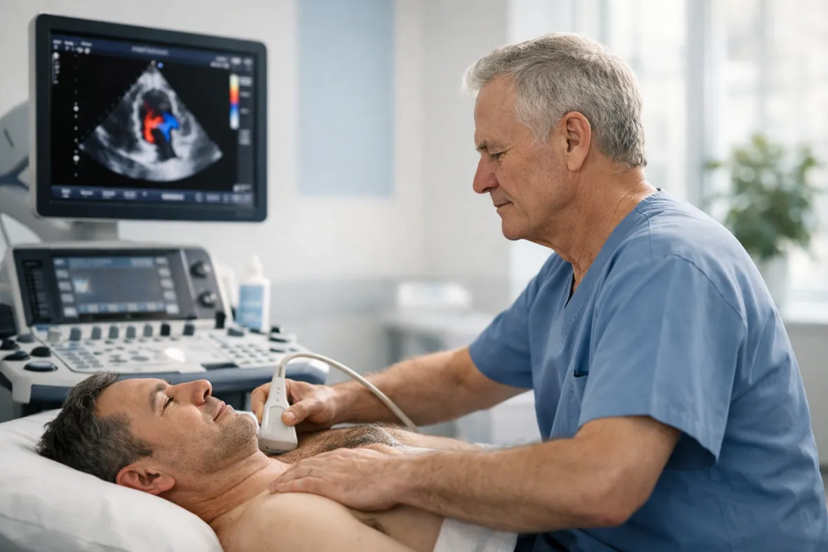
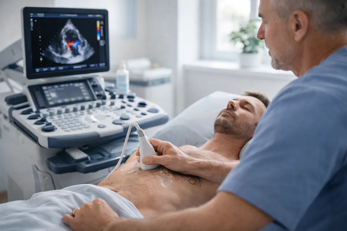
An ECG records the heart’s electrical activity (rhythm and conduction). An echo (echocardiogram) creates moving images of
the heart’s structure and pumping function (valves, chambers, muscle, Doppler blood flow).
If you have severe chest pain, collapse, severe breathlessness at rest, or stroke symptoms, seek urgent medical care first.
Private testing is best for clarity and next-step guidance — not emergency treatment.
Fast answer (the “right test first” shortcut)
You don’t need to memorise cardiology. Use this: ECG is the rhythm test. Echo is the structure/function test.
Many real-world pathways use both — but usually in a deliberate order.
ECG first
If your main symptom feels electrical
Palpitations, skipped beats, racing heart, episodic dizziness, fainting episodes, or “is my rhythm normal?” start with ECG (and often
longer rhythm monitoring if episodes come and go).
Job: capture rhythm and conduction patterns.
Reality: if symptoms are intermittent, a single ECG may be normal — monitoring may be needed.
Echo first
If your main symptom feels structural
Breathlessness on exertion, reduced exercise tolerance, a new murmur, valve concerns, or “is my heart pumping normally?” are echo questions.
Job: see chambers, valves, pumping function, Doppler blood flow.
Severe chest pain, collapse, severe breathlessness at rest, or stroke symptoms need urgent assessment (ECG + clinical pathway).
Don’t use a private scan to replace emergency care.
Echo vs ECG: side-by-side comparison
This table is designed for patients: plain-English differences, plus what each test cannot do (so you don’t book the wrong thing).
ECG is the fastest way to answer “is my heart rhythm normal right now?” It’s often the first step in urgent pathways (especially chest pain),
and a common first step for palpitations.
ECG-first symptom patterns
Palpitations (racing heart, skipping, fluttering), especially with dizziness or near-fainting.
Chest pain that is new, severe, or associated with sweating/nausea/shortness of breath — urgent pathway first.
Collapse or fainting (syncope), particularly during exertion — urgent clinical assessment is appropriate.
If your palpitations come and go
A normal ECG can still be “true” — it just means the rhythm was normal at that moment.
If symptoms are episodic, clinicians often use longer monitoring (Holter/event monitor) to catch the episode.
Echo may be added to check whether there’s a structural reason the rhythm is happening.
When an echo should come first
Echo answers: “How is my heart built, and how is it working?” It’s the main imaging test for murmurs/valves and a key test for
breathlessness where a cardiac contribution is being considered.
Echo-first symptom patterns
Heart murmur (new finding or follow-up): echo evaluates valve narrowing/leakage and haemodynamic significance.
Breathlessness on exertion or reduced exercise tolerance: echo assesses pumping function, valves and pericardial fluid.
Monitoring known conditions (valve disease, cardiomyopathy patterns) at the interval advised by your clinician.
When you may need both (common real-world pathways)
ECG and echo are complementary: one is electrical, one is mechanical/structural. Here are the scenarios where clinicians commonly combine them.
Palpitations + breathlessness
Capture rhythm + check structure
ECG (or monitoring) answers “what rhythm is it?” Echo answers “is there an underlying structural contributor (valves, cardiomyopathy pattern, function)?”
Murmur + symptoms
Valve assessment + baseline rhythm
Echo grades valve disease. ECG provides rhythm baseline and can show strain patterns that support the clinical picture.
Monitoring known diagnosis
Follow trends, not single numbers
Echo trends valve severity and function. ECG/monitoring checks rhythm evolution where clinically relevant.
Acute symptoms
Urgent pathway first
In acute chest pain or collapse pathways, ECG is immediate. Echo may follow in hospital assessment depending on findings and clinical need.
Best way to choose
Start with your “job to be done”: rhythm question → ECG. Structure/function question → echo.
If you’re unsure, use the symptom guide and then choose the test that answers your main concern.
Sonoworld provides private echocardiography (echo). ECG is commonly arranged via GP, cardiology, urgent care, or monitoring services
depending on your symptom urgency and pattern.
If you’re booking an echo
Book a private echocardiogram if your main concern is valves, pumping function, a murmur, breathlessness on exertion, or monitoring a known condition.
Bring any prior echo/ECG/Holter reports if you have them (comparisons make interpretation stronger).
Not really. Echo and ECG answer different questions. Echo images structure and pumping; ECG records rhythm and conduction.
Many pathways use both because they provide different types of information.
Will an echo show an arrhythmia?
Echo is not designed to capture rhythm episodes in the way an ECG does. It can sometimes show clues (for example, effects on function),
but rhythm diagnosis is an ECG/monitoring job.
Will an ECG show valve disease or how well my heart pumps?
ECG is not an imaging test. It can suggest strain patterns in some contexts, but it does not directly show valve structure or pumping function.
Echo is the test that images valves and function.
I have palpitations — should I book an echo?
Start with ECG/rhythm capture if the main question is rhythm. Echo is often added to check structure and function,
especially if palpitations are accompanied by breathlessness, dizziness, a murmur, or reduced exercise tolerance.
Is an echocardiogram safe?
A standard transthoracic echo uses ultrasound (sound waves), not radiation, and is generally well tolerated.
Read: Echo safety and risks.