A colour Doppler arterial duplex ultrasound-scan that evaluates blood flow, plaque, and vessel narrowing in the legs, arms, or neck. No preparation required. Same-day report. No GP referral needed.
Prefer to speak to us first? Call 020 3633 4902
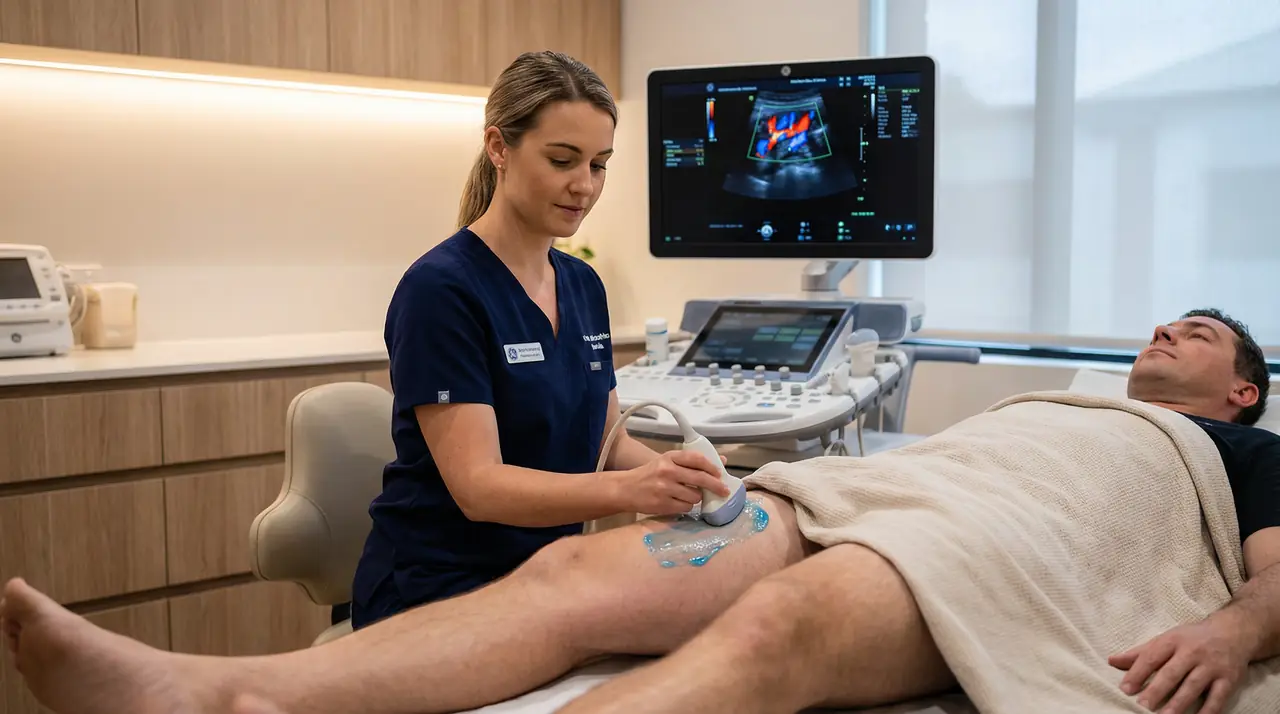
An arterial duplex ultrasound-scan combines two complementary techniques in a single examination. Greyscale B-mode imaging produces real-time cross-sectional pictures of the artery wall, revealing plaque deposits, calcification, and vessel diameter. Colour Doppler then maps the speed and direction of blood flow through the lumen, identifying areas of turbulence, stenosis, or occlusion that greyscale alone cannot detect.
The scan is performed by a HCPC-registered consultant sonographer using a GE Voluson system. It is non-invasive, painless, and requires no preparation — you can eat and drink normally beforehand and continue all medications as prescribed.
Peripheral arterial disease (PAD) affects approximately 20% of people over 60 in the UK, yet many remain undiagnosed until symptoms become disabling. An arterial duplex ultrasound-scan detects the haemodynamic changes that precede those symptoms, allowing timely intervention before the disease progresses.
Doppler colour flow — red and blue colour mapping on the screen represents blood moving towards and away from the probe. Areas of turbulent or high-velocity flow appear as colour aliasing, which the sonographer uses to grade the severity of any stenosis.
The scan can be performed on the lower limbs, upper limbs, or neck arteries. The table below shows the vessels examined and the conditions the duplex ultrasound-scan can identify in each region.
| Region | Vessels Examined | Conditions Detected |
|---|---|---|
| Lower limb | Common femoral, superficial femoral, popliteal, anterior & posterior tibial, peroneal arteries | PAD, stenosis, occlusion, aneurysm, graft surveillance |
| Upper limb | Subclavian, axillary, brachial, radial, ulnar arteries | Thoracic outlet syndrome, subclavian steal, stenosis, vasospasm |
| Carotid & neck | Common carotid, internal carotid, external carotid, vertebral arteries | Atherosclerotic plaque, carotid stenosis, intima-media thickening, dissection |
| Renal arteries | Main renal arteries, segmental branches | Renal artery stenosis, renovascular hypertension |
| Mesenteric | Coeliac axis, superior mesenteric artery | Mesenteric ischaemia, post-prandial abdominal pain |
Ankle-Brachial Index (ABI) — where clinically indicated, the sonographer can combine the duplex examination with ABI measurement to quantify the severity of lower limb arterial disease.
Arterial disease develops gradually and often silently. Many patients book this scan after noticing symptoms that their GP has attributed to other causes, or after a routine blood test reveals elevated cholesterol or blood glucose. The scan provides objective haemodynamic data that guides clinical decision-making.
Seek urgent medical attention if you experience:
These symptoms require immediate A&E assessment. Call 999 or go directly to your nearest emergency department.
The examination follows a structured protocol that covers greyscale imaging, colour Doppler mapping, and spectral waveform analysis. No preparation is required beforehand.
The sonographer takes a brief history of your symptoms, risk factors, and any previous vascular interventions. This guides the scan protocol and ensures the most relevant vessels are examined.
Ultrasound gel is applied to the skin and the probe is placed over each vessel in turn. The sonographer measures vessel diameter and assesses the arterial wall for plaque, calcification, and intima-media thickening.
Colour flow is applied to map blood movement through each vessel. Areas of turbulence or high-velocity flow indicate stenosis. The sonographer records peak systolic velocities and velocity ratios to grade any narrowing.
Pulsed-wave Doppler produces a spectral waveform at each sample site. The waveform shape — triphasic, biphasic, or monophasic — indicates the level of arterial disease distal to the measurement point.
The sonographer discusses the findings with you immediately after the scan. A full written report with annotated images is issued the same day and can be sent directly to your GP or referring consultant.
All prices are all-inclusive. There are no additional charges for the written report, annotated images, or same-day results.
Private medical insurance: Sonoworld is recognised by most major UK insurers including Bupa, AXA Health, Aviva, and Vitality. Please obtain an authorisation code from your insurer before booking and bring it to your appointment.
No GP referral is required. You can book directly through the Sonoworld website or by calling 020 3633 4902. If you do have a referral letter, please bring it to your appointment as it helps the sonographer tailor the examination to your clinical question.
A single-limb examination typically takes 25–35 minutes. A bilateral scan takes 45–60 minutes. Allow a little extra time for the clinical history and verbal feedback at the end of the appointment.
The scan is completely painless and non-invasive. The sonographer applies a small amount of water-based gel to the skin and moves the probe gently over the area. There is no radiation, no needles, and no contrast agents involved.
An arterial duplex scan examines the arteries — the vessels that carry oxygenated blood away from the heart. A venous duplex scan (such as a DVT scan) examines the veins, which return blood to the heart. The two scans assess different pathologies: arterial disease typically causes reduced blood flow and ischaemia, while venous disease causes obstruction and reflux. If you are unsure which scan you need, call the clinic and a member of the team will advise you.
The written report with annotated images is issued on the same day as your appointment. It is sent to you by secure email and can also be sent directly to your GP or referring consultant if you provide their contact details.
Yes. Arterial duplex ultrasound is the first-line imaging investigation for suspected PAD according to NICE guidelines (NG19). The scan identifies the location and severity of stenosis or occlusion, and the spectral waveform pattern indicates the haemodynamic significance of any disease found. This information directly informs decisions about conservative management, angioplasty, or surgical bypass.
Yes. Sonoworld is recognised by most major UK private medical insurers. Contact your insurer before booking to obtain an authorisation code, then bring that code to your appointment. If your insurer requires a GP or consultant referral letter, please arrange this in advance.
Sonoworld is a CQC-registered private diagnostic ultrasound clinic located in the heart of Marylebone, close to Harley Street. The clinic is easily accessible from Oxford Circus, Regent's Park, and Baker Street stations.