A bilateral duplex Doppler ultrasound-scan that assesses valve function and blood flow direction in the leg veins. Identifies venous insufficiency, maps reflux patterns, and supports treatment planning for varicose veins and venous skin changes. Written report within 24 hours. No GP referral required.
No GP referral needed
Report within 24 hours
Marylebone, Central London
HCPC Registered Sonographer
Healthy leg veins rely on a series of one-way valves to push blood back towards the heart against gravity. When those valves weaken or fail, blood pools in the vein and flows backwards — a process called venous reflux, or venous insufficiency. Over time, this raised venous pressure causes the familiar symptoms: aching, heaviness, ankle swelling, and, in more advanced cases, skin discolouration, eczema-like changes, and venous ulcers.
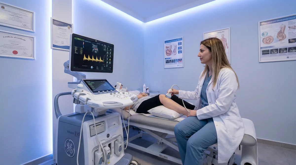
A venous reflux ultrasound-scan is a duplex Doppler examination that combines greyscale B-mode imaging with colour flow Doppler to map vein anatomy and detect valve incompetence. The sonographer uses gentle calf compression and release manoeuvres to provoke reflux and measure its duration and extent. Because venous disease is rarely confined to one limb, the assessment is always performed bilaterally — both legs are examined to give a complete clinical picture.
At Sonoworld, the examination is performed by an HCPC-registered vascular sonographer using a GE Voluson system. A written report is issued within 24 hours and is designed to be shared directly with your GP, vascular surgeon, or phlebologist.
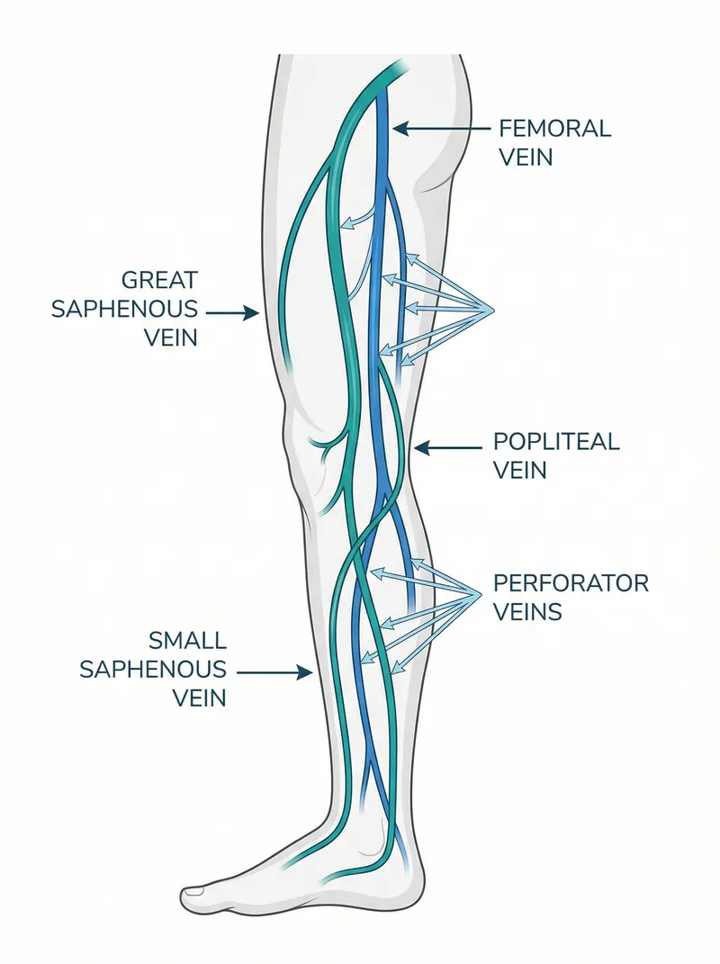
Vein systems assessed in this scan
Great saphenous vein (GSV) — the principal superficial vein; the most common site of reflux driving varicose veins
Small saphenous vein (SSV) — the posterior superficial vein; assessed at the sapheno-popliteal junction
Deep veins (femoral, popliteal) — assessed for patency and any deep reflux contribution
Perforator veins — where clinically relevant, particularly in skin change or ulcer assessment
Sapheno-femoral junction (SFJ) — key reflux entry point at the groin
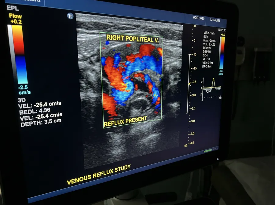
Colour Doppler flow — direction, duration, and velocity of reflux using compression manoeuvres
Reflux duration thresholds
Reflux is defined by the duration of retrograde flow following a standardised compression manoeuvre. The accepted clinical thresholds are:
Superficial veins (GSV, SSV)> 0.5 seconds
Deep veins (femoral, popliteal)> 1.0 second
Perforator veins> 0.35 seconds
Clinical Assessment
What the Venous Reflux Ultrasound-Scan Detects
The examination uses greyscale imaging and colour Doppler to assess vein anatomy, valve competence, and flow direction throughout the superficial and deep venous systems of both legs. The table below summarises the structures assessed and the conditions identified.
Structure
Assessment Method
Conditions Identified
Great saphenous vein (GSV)
Greyscale diameter; Doppler reflux duration at SFJ and along the thigh and calf
Venous disease is classified using the internationally recognised CEAP system. A reflux ultrasound-scan provides the objective evidence needed to assign the correct clinical class and plan appropriate management.
C0–C1: No visible disease / telangiectasiaReassurance
Venous insufficiency is frequently bilateral even when symptoms appear unilateral. Assessing only the symptomatic leg misses clinically significant disease in the other limb and produces an incomplete picture for treatment planning. All venous reflux studies at Sonoworld are performed on both legs as standard.
Indications
When a Venous Reflux Ultrasound-Scan Makes Sense
People book this scan because their legs feel different, look different, or because they are planning treatment and need an objective map of what is happening. The scan answers one core question: are the vein valves working properly, and if not, where is the reflux originating?
Aching, heaviness, or fatigue in the legs — worse as the day progresses or after prolonged standing
Visible varicose veins — bulging, rope-like veins on the thigh, calf, or behind the knee
Ankle swelling that improves with leg elevation or compression stockings
Itching, tightness, or a burning sensation around the lower leg or ankle
Skin discolouration, lipodermatosclerosis, or eczema-like changes around the ankle
A poorly healing wound or venous ulcer on the lower leg
Pre-treatment mapping before foam sclerotherapy, endovenous laser ablation (EVLA), or radiofrequency ablation (RFA)
Follow-up after varicose vein treatment to confirm closure and assess for recurrence
Not the right scan for sudden swelling or suspected DVT
Sudden one-sided leg swelling, calf pain, redness, or warmth — particularly with shortness of breath — requires urgent medical assessment and a DVT ultrasound-scan pathway, not a reflux study. If in doubt, seek urgent care first.
What the scan gives you
Clarity: turns "I think it's varicose veins" into a documented finding — which veins, where, and how severe
Direction: a structured report that supports conservative management, specialist referral, or treatment planning
Reassurance: if reflux is not demonstrated, you can stop guessing and explore alternative explanations with confidence
Treatment map: vascular clinicians need a reflux map before performing EVLA, RFA, or foam sclerotherapy — this scan provides exactly that
Preparation for a venous reflux ultrasound-scan is straightforward. There is no fasting, no special diet, and no need to adjust medications. A few simple steps ensure the scan can be performed efficiently and comfortably.
Clothing
Wear or bring loose clothing that allows easy access above the knee. Shorts or loose trousers are ideal. Compression stockings should be removed before the scan — you can bring them with you and put them back on afterwards.
Skin preparation
Avoid applying lotions, creams, or oils to your legs on the day of the scan. These can interfere with ultrasound gel contact and reduce image quality. Clean, dry skin gives the best results.
Hydration and diet
Drink water normally before the appointment — good hydration supports venous filling and improves image quality. No fasting is required. Continue eating and drinking as usual.
Medications
Continue all medications as prescribed. If you take anticoagulants (e.g., warfarin, apixaban, rivaroxaban), there is no need to stop them for this scan. Bring a list of your current medications to the appointment.
What to bring
Insurance authorisation code (if applicable)
Any previous venous duplex or vascular reports
List of current medications
GP or specialist referral letter (if you have one — not required)
Comfort note
The scan is non-invasive. You will feel only cool gel and gentle probe pressure on your legs. For part of the examination you may be asked to stand or adopt a semi-standing position — this is normal and helps to demonstrate reflux under gravitational load.
If any area of your leg is tender or has an open wound, tell the sonographer before the scan begins. The technique can be adjusted accordingly.
Your Appointment
What Happens During Your Venous Reflux Ultrasound-Scan
The appointment is structured to be calm and unhurried. Both legs are assessed in sequence, with the sonographer explaining findings as the scan progresses. Here is what to expect at each stage.
1
Clinical history
The sonographer confirms your symptoms, duration, any prior vein history or treatment, and your primary goal — whether that is reassurance, diagnosis, or pre-treatment mapping. You sign a consent form before the scan begins.
2
Greyscale vein mapping
Both legs are exposed and ultrasound gel is applied. The sonographer maps the superficial and deep vein anatomy in greyscale, assessing vein calibre, wall morphology, and compressibility from groin to ankle.
3
Colour Doppler reflux assessment
Colour Doppler is applied at each key junction (SFJ, SPJ, perforators). Gentle calf compression and release manoeuvres provoke reflux. Duration and extent of retrograde flow are measured and recorded.
4
Standing assessment
Where clinically indicated, you will be asked to stand or adopt a semi-standing position. Gravity increases venous pressure and makes reflux easier to demonstrate and measure accurately.
5
Verbal feedback & report
The sonographer explains findings in plain English at the end of the scan — which veins reflux, where, and what that means for your next steps. A written report is issued within 24 hours and can be shared with your GP or vascular specialist.
Online Booking
Book Your Venous Reflux Ultrasound-Scan
Choose your preferred date and time below. Booking is confirmed immediately. No GP referral is required. Both legs are always assessed as part of the bilateral study.
Price includes bilateral assessment, verbal feedback, and written report within 24 hours. Insurance patients: please bring your authorisation code.
Preparation reminder
No lotions or creams on legs on the day
Wear loose clothing — access above the knee needed
Remove compression stockings before the scan
Continue all medications as normal
Drink water normally — no fasting required
What happens after the scan
Verbal feedback given immediately after the scan
Written report with reflux findings issued within 24 hours
Report designed to share with GP, phlebologist, or vascular surgeon
Follow-up appointment available if surveillance or post-treatment review is needed
Transparent Pricing
Venous Reflux Scan Price
One fixed price. Venous insufficiency is always assessed bilaterally — both legs are examined as standard to ensure a complete clinical picture. No hidden fees. No GP referral required.
Insurance patients are welcome. Please bring your authorisation code. Sonoworld is recognised by most major insurers including Bupa, AXA Health, Aviva, and Vitality.
Frequently Asked Questions
Venous Reflux Scan — Common Questions
Is this the same as a DVT ultrasound-scan?
No. A DVT scan looks for acute blood clots (thrombus) in the deep veins and is the appropriate test for sudden, painful, one-sided leg swelling. A venous reflux scan assesses valve function and chronic venous insufficiency — it is the correct test for varicose veins, aching legs, ankle swelling, and skin changes that have developed gradually. If you have sudden swelling with pain or redness, please seek urgent care and consider a DVT scan instead.
Do I need a GP referral?
No. You can book a venous reflux ultrasound-scan at Sonoworld directly, without a GP referral. The scan is available to anyone who wishes to assess their venous health, whether for reassurance, diagnosis, or pre-treatment planning. If you have a referral letter, bring it along — it provides useful clinical context.
Why are both legs always scanned?
Venous insufficiency is frequently bilateral even when symptoms appear to affect only one leg. Assessing only the symptomatic limb misses clinically significant disease in the other and produces an incomplete picture for treatment planning. Vascular surgeons and phlebologists require bilateral data before planning interventions such as EVLA, RFA, or foam sclerotherapy. For this reason, all venous reflux studies at Sonoworld are bilateral as standard.
Is the scan safe?
Yes. Ultrasound-scan uses sound waves, not radiation. It is safe for all patients, including those who are pregnant or taking anticoagulant medication. The examination is non-invasive — there are no needles, no injections, and no internal examination. You will feel only cool gel and gentle probe pressure on your legs.
How long does the scan take?
A bilateral venous reflux study typically takes 30–45 minutes, depending on clinical complexity. Both legs are assessed in sequence. Your booking confirmation will include the allocated appointment time. The appointment is designed to be unhurried — the sonographer will take time to explain findings clearly before you leave.
Will you tell me what to do next?
Yes. The sonographer provides verbal feedback at the end of the scan, explaining what was found and what it means. The written report, issued within 24 hours, includes a structured clinical summary and next-step guidance designed to be shared with your GP or specialist. If reflux is present, the report documents which veins are affected and the severity — the information a vascular clinician needs to plan conservative or interventional management.
Can I cancel or reschedule my appointment?
Yes. Appointments can be cancelled or rescheduled up to 24 hours before the appointment time without charge. Cancellations made within 24 hours may be subject to a cancellation fee. Please call 020 3633 4902 or email info@sonoworld.co.uk to make changes.
Find Us
Sonoworld — Marylebone, London
Clinic address
29 Weymouth Street London W1G 7DB
Nearest stations: Regent's Park (Bakerloo) · Great Portland Street (Circle, H&C, Metropolitan) · Baker Street (Jubilee, Bakerloo, Metropolitan)