Ultrasound-Guided Injections: Why Guidance Matters
By Daniela Stan, Consultant Sonographer, MSc · 29 Weymouth Street, Marylebone · Medically reviewed by Daniela Stan, Consultant Sonographer, MSc — 25 July 2026 · Last reviewed: 31 July 2026
An injection only works if it reaches the structure causing the pain. Placed by feel using surface landmarks, a meaningful proportion of injections end up near the target rather than in it — which is one reason injections fail. Ultrasound shows the needle and the target together in real time, so placement is watched rather than assumed.
What an ultrasound-guided injection is
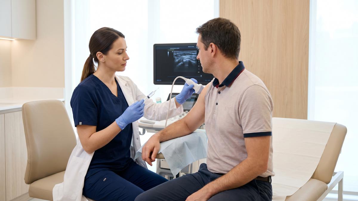
The area is scanned first to confirm the diagnosis and identify the target. The skin is cleaned, and the needle is advanced under direct ultrasound vision — the operator watches the needle tip travel to the intended structure and sees the injected fluid spread into it.
That last detail is the part people underestimate. Seeing the medication distribute within the target confirms delivery. Without imaging, there is no way to know whether it went where it was intended.
Why guidance matters
Landmark-guided injection relies on the clinician estimating the position of a structure from the surface anatomy. That works reasonably well for large, superficial joints and much less well for small compartments, deep bursae and tendon sheaths.
Published comparisons consistently find that image guidance improves the accuracy of needle placement. Across a range of joints, ultrasound-guided injections were accurate in 83 per cent of cases compared with 66 per cent for injections guided by clinical examination alone. The margin varies considerably by site:
| Target |
Ultrasound-guided |
Landmark-guided |
| Biceps tendon sheath |
86.7% |
26.7% |
| Acromioclavicular joint |
93.6% |
68.2% |
| Glenohumeral joint |
92.5% |
72.5% |
| Knee joint |
95–96% |
78–83% |
| Subacromial space |
No significant accuracy difference — but better pain and function outcomes at six weeks |
The pattern is consistent: accuracy matters most where the target is small, deep, or surrounded by structures you would rather not inject — the biceps tendon sheath being the clearest example, where landmark placement misses roughly three times out of four. For the large subacromial space, accuracy is comparable, yet guided injections still produced greater reduction in pain and improvement in function at six weeks.
It also matters for what should not be injected. Ultrasound can show a tendon that is torn rather than inflamed, where a corticosteroid injection would be the wrong treatment.
Types of injection
- Corticosteroid — the most common. Reduces inflammation in a joint, bursa or tendon sheath. Relief typically develops over several days and can last weeks to months.
- Local anaesthetic — often combined with steroid for immediate relief, and sometimes used diagnostically: if numbing a specific structure abolishes the pain, that structure is confirmed as the source.
- Barbotage (needling and aspiration) — used for calcific tendinopathy, where the calcium deposit is broken up and aspirated.
- Aspiration — drawing off fluid from a joint, bursa or ganglion, both to relieve pressure and to send fluid for analysis.
- Hydrodistension — injecting volume into a joint capsule, used in frozen shoulder.
Conditions treated
| Area |
Commonly injected |
| Shoulder |
Subacromial bursitis, calcific tendinopathy, acromioclavicular joint, frozen shoulder |
| Elbow |
Tennis and golfer’s elbow, olecranon bursitis |
| Wrist and hand |
Carpal tunnel, De Quervain’s, trigger finger, thumb base arthritis, ganglion aspiration |
| Hip |
Trochanteric bursitis, gluteal tendinopathy, iliopsoas bursa, hip joint |
| Knee |
Knee joint, pes anserine bursitis, Baker’s cyst aspiration |
| Foot and ankle |
Plantar fascia, ankle joint, Morton’s neuroma |
Injection into the Achilles tendon itself is generally avoided because of the risk of weakening it — see Achilles tendon pain.
What to expect, and afterwards
The appointment takes around 30 minutes including the diagnostic scan. Most people describe pressure rather than sharp pain. You can usually drive afterwards, though not if a large joint in your driving leg has been injected.
In the first 24 to 48 hours some people experience a temporary increase in pain as the local anaesthetic wears off before the steroid takes effect. This is common and settles. Rest the area for the first day or two.
Over the following weeks the anti-inflammatory effect builds. This is the window in which to do the rehabilitation — an injection that reduces pain without any change in loading tends to be followed by the same problem returning.
An injection is rarely a treatment on its own. It is most effective as a way of making rehabilitation possible.
Risks and limits
- Post-injection flare — temporary increase in pain, usually settling within 48 hours
- Skin changes — thinning or lightening at the injection site, more likely with superficial injections
- Raised blood sugar for a few days, which matters if you have diabetes
- Tendon weakening if steroid is placed into a tendon rather than around it — a specific reason guidance matters
- Infection — rare, but any hot, swollen, increasingly painful joint with fever after an injection needs same-day medical assessment
There are limits on repetition. Injections into the same site are not repeated indefinitely, and if two well-placed injections have not helped, the answer is usually a different treatment rather than a third.
Seek same-day medical assessment if, after an injection, you develop: a hot, swollen, increasingly painful joint; fever or feeling generally unwell; spreading redness; or pain that is worsening after 48 hours rather than settling.
From our practice
From our practice
Calcific tendinopathy of the shoulder is one of the conditions where ultrasound-guided barbotage — needling and aspiration of the calcium deposit — produces the most dramatic results.
Patients who have had months of severe shoulder pain, often worse than a rotator cuff tear, can experience near-complete resolution of symptoms within two to four weeks of a single ultrasound-guided barbotage procedure. The key is accurate needle placement within the calcium deposit, which is only reliably achievable under ultrasound guidance.
Describes general patterns across the patients we treat, not any individual case. Outcomes vary and suitability is assessed individually.
Common questions
Is an ultrasound-guided injection painful?
Most people describe pressure rather than sharp pain. Local anaesthetic is usually included, so the area often feels numb immediately afterwards. Some discomfort as that wears off is normal.
How long does a cortisone injection last?
It varies with the condition and the structure injected. Relief commonly lasts several weeks to several months. It works best when used to create a window for rehabilitation rather than as a standalone treatment.
Do I need a scan before having an injection?
Yes, and it is done in the same appointment. The scan confirms the diagnosis and identifies the target — and occasionally shows that an injection is not the right treatment, for example where a tendon is torn rather than inflamed.
How soon will it work?
Local anaesthetic gives immediate but temporary relief. The steroid effect typically develops over several days to two weeks. A temporary flare in the first 24 to 48 hours is common.
How many injections can I have?
Injections into the same site are not repeated indefinitely. If two accurately placed injections have not produced lasting benefit, a different approach is usually more appropriate than a third.
How much does an ultrasound-guided injection cost?
Ultrasound-guided injections start from £250, which includes the diagnostic scan. You receive instant verbal results at the appointment, with the written report following within 24 hours.
Related: tennis and golfer’s elbow, wrist and hand pain, shoulder pain, and our guide to MSK ultrasound.
References
- Aly AR, Rajasekaran S, Ashworth N. Ultrasound-guided shoulder girdle injections are more accurate and more effective than landmark-guided injections: a systematic review and meta-analysis. British Journal of Sports Medicine 2015;49(16):1042–9.
- Ultrasound-guided infiltrative therapy of the knee: accuracy, clinical outcomes and cost-effectiveness. Journal of Medical Imaging and Interventional Radiology 2025.
- Sibbitt WL et al. Does ultrasound guidance improve the outcomes of arthrocentesis and corticosteroid injection of the knee? Scandinavian Journal of Rheumatology 2012;41:66–72.
- Soh E et al. Image-guided versus blind corticosteroid injections in adults with shoulder pain: a systematic review. BMC Musculoskeletal Disorders 2011;12:137.
CQC-registered clinic · BMUS member · ICO-registered. Reports can be shared securely with your NHS GP or specialist at your request.
Our injection service: ultrasound-guided cortisone injections from £250, including the diagnostic scan. See also all MSK ultrasound scans.