- Home
- Scans for Women
- Scans for Men
- Msk
- Pregnancy
- Cardiovascular
- About
- Book a Scan
- Blog
A hernia occurs when an internal part of the body pushes through a weakness in the muscle or surrounding tissue wall. In many cases, hernias cause no symptoms or very few symptoms. You may notice a swelling or lump in your tummy (abdomen) or groin that can often be pushed back in or will disappear when you lie down. Coughing or straining may make the lump to re-appear.
Types of hernias:
Hernias can occur throughout the body, but most often develop in the areas of your body between your chest and hips.

The cost of this ultrasound scan is only £164.
Types of hernias:
Hernias can occur throughout the body, but most often develop in the areas of your body between your chest and hips.
Some of the more common types of hernia are:
Inguinal hernias occur when fatty tissue or a part of your bowel pokes through into your groin at the top of your inner thigh. This is the most common type of hernia, and it mainly affects men. It is often associated with ageing and repeated strain on the abdomen.
Femoral hernias also occur when fatty tissue or a part of your bowel pokes through into your groin at the top of your inner thigh. They are much less common than inguinal hernias and tend to affect more women than men. Like inguinal hernias, femoral hernias are also associated with ageing and repeated strain on the abdomen.
Umbilical hernias occur when fatty tissue or a part of your bowel pokes through your abdomen near your belly button (navel).
Other types of hernia that can affect the abdomen:
The hernia ultrasound scan includes evaluation of either:
No preparation is necessary for this scan.
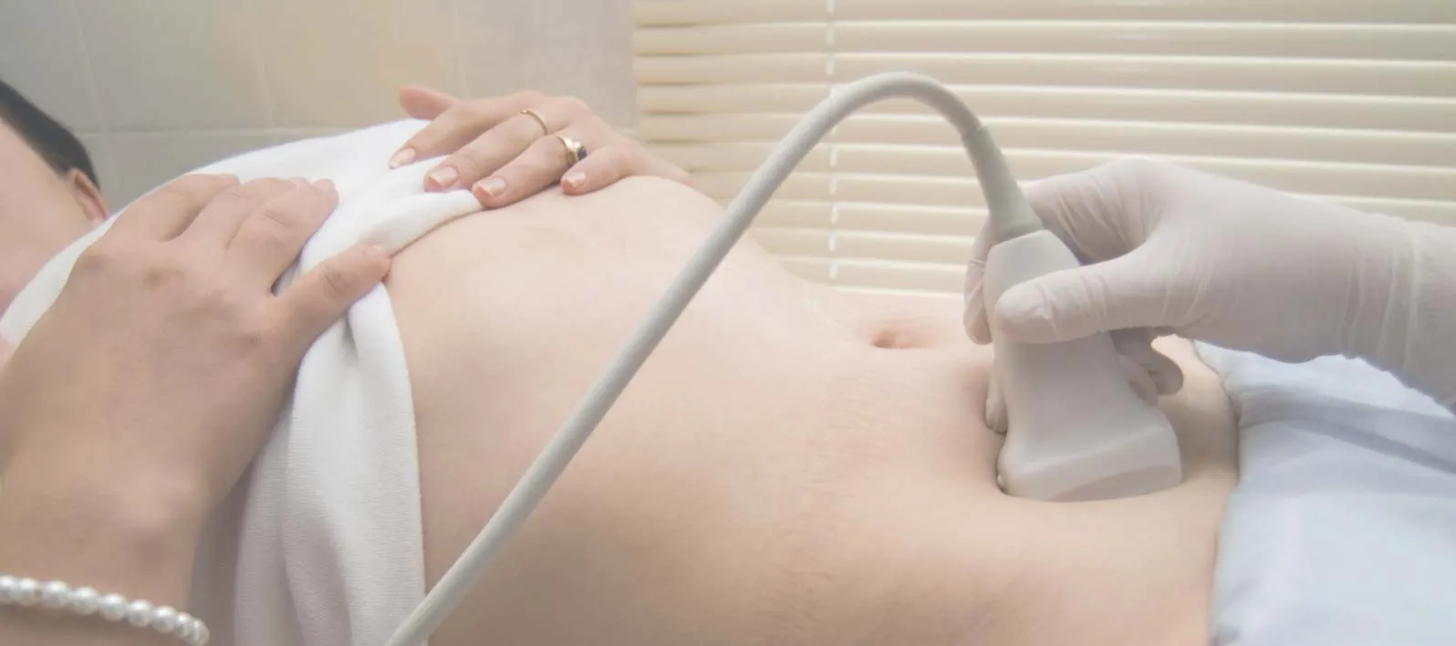
Before the scan, our sonographer will explain the examination procedure. You will be asked to lie on the examination couch, exposed part of your abdomen. We will put a little gel on your skin and a small ultrasound probe will be used to obtain images of your internal organs.
During and after the examination, our sonographer will explain the findings and an ultrasound report will be issued to take away with you.

Ultrasound imaging is a medical diagnostic technique where sound waves are being used to image various parts of the body.
Other terms for ultrasound imaging are sonograms, US and sonography.
Ultrasound is widely used these days as it is painless and safe for adults, children and foetuses. There are no side effects such as the ones associated with radiation.
During the ultrasound scan, the sonographer rests a small probe over the skin. This probe produces sound waves, i.e. pulsations, that travel through the tissues. Some sound waves are being reflected back to the transducer, and the computer analyses the returning echoes and produces the image on the screen. It is the same principle as the sonar the navy uses.
Ultrasound is being used to image mostly solid organs such as liver, kidneys, uterus and ovaries, muscles and blood vessels and babies in the womb.
It has, however, limited value in organs such as lungs, bone, stomach and bowel/colon.
Ultrasound images are black and white but colour Doppler is being used to evaluate organ and blood vessel blood flow and this is what the red and blue colours on the screen are.
In our ultrasound scan clinic in Harley Street, you can book a private abdominal scan without the need for a doctor's referral.