
- Home
- Women
- Men
- MSK
- Cardiovascular
- Screening
- About
- Book a Scan
- Blog
A private echocardiogram or Echo is a non-invasive heart ultrasound that images the chambers, valves, and blood flow of your heart in real time. Performed in Marylebone by BSE-accredited consultant cardiac sonographers, from £350, with a written report within 24 hours. No GP referral required.
See what's included in our echocardiogram cost, how an echo compares to an ECG or cardiac MRI, or learn about stress echocardiography and transoesophageal echocardiography (TOE).

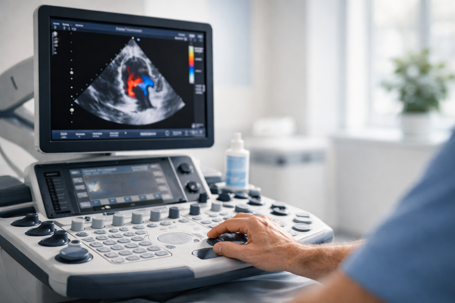
An echocardiogram (often called an "echo") uses high-frequency sound waves to create real-time moving images of your heart. It is the primary heart imaging tool for assessing heart structure and function — showing how the chambers fill and empty, how the valves open and close, and how blood flows through the heart with every beat.
At Sonoworld, every echocardiogram is performed by a consultant cardiac sonographer with specialist post-graduate training in cardiac ultrasound. Our team brings over 20 years of combined NHS and private cardiac imaging experience. The scan uses no radiation and requires no contrast dye or fasting — it is entirely non-invasive.
A written report — including measurements, Doppler findings, and clinical interpretation — is typically issued within 24 hours and can be shared directly with your GP, cardiologist, or referring clinician.

Every echo at Sonoworld is performed by a consultant cardiac sonographer — not a general ultrasonographer.
Echocardiography is the first-line imaging test for many cardiac conditions. The table below summarises the structures assessed and the clinical findings the scan can identify or monitor.
| Structure / Parameter | Assessment Method | Conditions Identified |
|---|---|---|
| Left ventricle (LV) | 2D dimensions; M-mode; ejection fraction (biplane Simpson) | Dilated cardiomyopathy, LV systolic dysfunction, hypertrophic cardiomyopathy |
| Right ventricle (RV) | 2D size; TAPSE; tricuspid annular motion | RV dilatation, pulmonary hypertension, RV dysfunction |
| Aortic valve | 2D morphology; Doppler gradients; planimetry | Aortic stenosis, aortic regurgitation, bicuspid aortic valve |
| Mitral valve | 2D leaflet motion; colour Doppler; pressure half-time | Mitral regurgitation, mitral stenosis, mitral valve prolapse |
| Tricuspid & pulmonary valves | Colour Doppler; spectral Doppler gradients | Tricuspid regurgitation, pulmonary hypertension estimation |
| Left atrium (LA) | 2D volume; LA size index | LA dilatation (associated with AF, diastolic dysfunction) |
| Pericardium | 2D circumferential assessment | Pericardial effusion, cardiac tamponade, constrictive pericarditis |
| Aortic root & ascending aorta | Parasternal long-axis measurement | Aortic root dilatation, aortic aneurysm screening |
Echo is the primary test for assessing valve stenosis (narrowing) and regurgitation (leakage). It measures severity grade and helps cardiologists decide whether to monitor, treat medically, or refer for surgical repair or replacement.
Echo measures ejection fraction (EF) — the percentage of blood pumped per beat. Normal EF is 55–70%. Below 40% suggests reduced function. Serial echos track whether treatment is working, in line with NICE NG106 guidance.
While AF is diagnosed by ECG or Holter monitor, an echo is often requested alongside to check for underlying structural causes — enlarged left atrium, valve disease, or impaired ventricular function.
Long-standing high blood pressure can cause the heart muscle to thicken (left ventricular hypertrophy). Echo detects and measures this thickening, influencing treatment decisions as outlined in NICE NG136.
Some structural differences (bicuspid aortic valve, atrial septal defect) are first detected in adulthood. Pericardial effusion and cardiac tamponade can also be identified and assessed for haemodynamic significance.
Most patients book privately because they want reassurance or faster answers than the NHS pathway can provide. An echocardiogram is used to investigate symptoms, assess a known diagnosis, or provide a cardiac baseline before exercise programmes or surgery.
Seek urgent care — do not book a private scan
Severe crushing chest pain, sudden severe breathlessness at rest, loss of consciousness, or new stroke-like symptoms (FAST) require immediate A&E attendance or call 999. A private echocardiogram is for assessment and monitoring — not for emergencies.
These tests answer different questions about your heart. They are often complementary and can be combined for a more complete cardiac assessment.
| Test | Best for |
|---|---|
| Echocardiogram | Heart structure, valve function, pumping strength |
| ECG | Heart rhythm, electrical conduction, AF detection |
| Holter monitor | Intermittent palpitations, 24–72 hr rhythm recording |
| Stress echo | Heart function under physical or pharmacological stress; coronary artery disease assessment |
| Transoesophageal echocardiogram (TOE) | Detailed valve and structural imaging where transthoracic views are limited; clot detection before cardioversion |
For a full side-by-side, read echo vs ECG vs cardiac MRI vs CT. Not sure which test fits your symptoms? Call us on 020 3633 4902.
For a standard transthoracic echocardiogram, there is no special preparation. You do not need to fast, and you can eat, drink, and take your usual medications normally.
Wear comfortable clothing that is easy to remove from the upper body. You will undress from the waist up and lie on a couch for the scan. A gown is provided.
Continue all medications as prescribed. There is no need to stop any medication for a standard transthoracic echo. Bring a list of your current medications (or photographs of the labels).
No fasting required for a standard transthoracic echo — eat, drink, and take your usual medications normally. Stress echo and transoesophageal echo (TOE) have different preparation requirements; if you are booked for one of these, you will receive separate instructions.
The scan is non-invasive. You will feel only cool gel and gentle probe pressure on your chest. You may be asked to breathe in, breathe out, or hold your breath briefly — this is normal and helps capture clear images.
We accept most major private medical insurers including Bupa, AXA Health, Aviva, and Vitality. Before your appointment:
The appointment is calm and unhurried. You will be guided step-by-step, with time to ask questions before, during, and after the scan. Here is what to expect at each stage.
We confirm your details, symptoms, medications, and any relevant history. If you have prior ECG, Holter, or echo reports, bring them — they help the sonographer focus the assessment. You sign a consent form before the scan begins.
You undress from the waist up and lie on your left side on the couch. Ultrasound gel is applied and the probe is placed on your chest in several positions to capture views of the heart chambers, valves, and blood flow.
Colour and spectral Doppler are used to measure blood flow velocity, direction, and turbulence through each valve and chamber. You may be asked to breathe in, breathe out, or hold your breath briefly to improve image quality.
After the scan, the sonographer shares initial observations in plain English. This is not a formal report but gives you an immediate sense of whether anything concerning has been seen and what the next steps are likely to be.
Your formal written report — including measurements, Doppler findings, and clinical interpretation — is typically delivered within 24 hours. It includes clear next-step guidance and can be sent directly to your GP or referring clinician.

Appointments are available Monday to Sunday. No GP referral is required. Same-day appointments are often available. Booking is confirmed immediately.
Choose your preferred date and time on our secure booking page. Confirmation is sent immediately by email. No GP referral required.
Price includes the scan, verbal feedback, and written report within 24 hours. Insurance patients: please bring your authorisation code.
One fixed price that includes the scan, verbal feedback, and written report. No GP referral required. Insurance accepted — please check your policy and obtain authorisation where required.
Insurance patients are welcome. Please bring your authorisation code. Sonoworld is recognised by most major insurers including Bupa, AXA Health, Aviva, and Vitality. See the full echocardiogram cost breakdown or our complete price list for all cardiac scans. After your scan, our guide to understanding your echo report explains every measurement in plain English.
This echocardiogram can be combined with one of our blood test packages for a more complete picture of your cardiovascular health. Pairing cardiac imaging with relevant blood markers — such as cholesterol, BNP, or inflammatory markers — gives you and your clinician a fuller assessment in a single visit.
No. A transthoracic echocardiogram is completely non-invasive. You will feel only cool ultrasound gel and gentle probe pressure on your chest. There are no needles, no injections, and no internal examination. Most patients find the scan straightforward and comfortable.
A standard transthoracic echocardiogram typically takes 30–45 minutes, depending on clinical complexity. This includes the clinical history, the scan itself, and the preliminary discussion at the end. Your appointment is never rushed — the sonographer will take time to explain findings clearly before you leave.
No. You can book a private echocardiogram at Sonoworld directly, without a GP referral. The scan is available to anyone who wishes to assess their cardiac health. If you have a referral letter from your GP or cardiologist, bring it along — it provides useful clinical context and may be required by your insurer.
An echocardiogram uses ultrasound to create moving images of the heart's structure — showing chambers, valves, and blood flow. An ECG (electrocardiogram) records the heart's electrical activity and is used to assess rhythm and conduction. They answer different clinical questions and are often complementary. If your concern is primarily about palpitations or irregular rhythm, an ECG or Holter monitor may be the more appropriate first test. If you are unsure, call us and we will guide you.
Ejection fraction (EF) is the percentage of blood pumped from your left ventricle with each heartbeat. Standard interpretation thresholds: 55–70% normal, 40–54% mildly reduced, below 40% reduced (possible heart failure), above 70% may indicate hypertrophic cardiomyopathy. Your echo report will state your EF and explain it in plain English. For a full walkthrough of what EF means clinically, see our dedicated guide on ejection fraction explained.
Yes. A transthoracic echocardiogram uses ultrasound — not magnetic fields or radiation — and is completely safe with pacemakers and implantable defibrillators (ICDs). Please let us know at the time of booking and mention it again when you arrive, so the sonographer can note it in your clinical history.
Yes. The scan is performed to the same BSE (British Society of Echocardiography) minimum dataset standards used in NHS cardiac departments. Our sonographers are HCPC-registered and BSE-accredited — the same professional standards required in NHS practice. The key differences are speed of access, a calm private environment, and a written report delivered within 24 hours rather than weeks.
You will receive verbal feedback from the sonographer immediately after the scan. A formal written report — including measurements, Doppler findings, and clinical interpretation — is typically issued within 24 hours. The report includes clear next-step guidance: reassurance, GP follow-up recommendation, or specialist cardiology referral if needed.
Yes. With your consent, we can send a copy of the report directly to your GP, cardiologist, or any other referring clinician. Please provide their contact details at the time of your appointment. You will always receive your own copy of the report regardless.
An echocardiogram assesses heart structure and function but does not directly image the coronary arteries (the blood vessels that supply the heart muscle). Coronary artery disease — the cause of most heart attacks — requires a different test such as CT coronary angiography or stress testing. If your concern is chest pain on exertion or suspected coronary artery disease, discuss this with your GP or cardiologist, who can direct you to the appropriate investigation. An echo is also not a substitute for an ECG or Holter monitor if your primary concern is heart rhythm.
Yes. Appointments can be cancelled or rescheduled up to 24 hours before the appointment time without charge. Cancellations made within 24 hours may be subject to a cancellation fee. Please call 020 3633 4902 or email [email protected] to make changes.
29 Weymouth Street
London W1G 7DB
Nearest stations: Regent's Park (Bakerloo) · Great Portland Street (Circle, H&C, Metropolitan) · Baker Street (Jubilee, Bakerloo, Metropolitan)
Open in Google MapsEchocardiography focuses on heart structure. If your concern involves heart rhythm, circulation, or stroke risk, one of these tests may complement your echo.
Heart rhythm & electrical conduction. 5–10 min.
24–72 hr continuous rhythm recording.
Plaque & narrowing in carotid arteries.
Deep vein clot assessment — leg swelling.
Symptom guides, condition explainers, and follow-up reading from our cardiovascular patient library.
What a murmur actually is, when it matters, and which echo findings rule things in or out.
Why palpitations happen, when an echo is appropriate, and when an ECG or Holter is the better first step.
A walkthrough of the valve, muscle, and structural conditions an echocardiogram can identify or monitor.
Plain-English introduction to how cardiac ultrasound works and what the different echo views show.
What EF means, how it's measured, and how to read the number on your echo report.


