
Home u203a Blog u203a Women's Health
Transvaginal Ultrasound u2014 Your Questions Answered
A transvaginal ultrasound is one of the most common scans in women's health u2014 and one of the most quietly worried-about. This guide answers the questions patients actually ask: why it is requested, exactly what happens in the room, whether it hurts, what is normal afterwards, and what a "bad" result really means.
Before anything else: two situations that should not wait for a booked scan
- Sudden, severe pelvic pain on one side u2014 especially with nausea, vomiting or feeling faint u2014 go to A&E or call 999. This can be a twisted ovary or a ruptured cyst.
- Pelvic pain or bleeding when you are, or could be, pregnant u2014 an ectopic pregnancy must be excluded urgently. Seek same-day medical review through your GP, NHS 111 or your nearest early pregnancy unit; do not wait for a routine appointment.
What are the reasons that doctors request a pelvic ultrasound?
The most common reasons for a pelvic ultrasound examination are:
- Pelvic pain u2014 if you are still deciding whether your pain warrants a scan at all, start with our guide to pelvic pain and when to get checked.
- Abnormal uterine or vaginal bleeding u2014 including any bleeding after menopause, which has its own rules and its own guide.
- Checking the status of an early pregnancy, including its location and heartbeat.
- Assessing the ovaries u2014 cysts, follicle tracking during fertility treatment, or surveillance where there is a strong family history of ovarian cancer or a known BRCA gene alteration.
- Investigating suspected fibroids, polyps or endometriosis-related changes.
On the NHS, the scan is requested by your doctor. In a private clinic such as Sonoworld you can self-refer u2014 your symptoms are the clinical indication, and no GP letter is needed.
The Pelvic Ultrasound Procedure: Transabdominal and Transvaginal
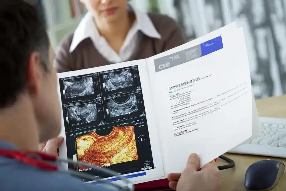
A pelvic ultrasound normally has two parts, and understanding both takes most of the anxiety out of the appointment.
Part one: the transabdominal scan. You will usually be asked to arrive with a full bladder u2014 your uterus sits behind your bladder, and a full bladder lifts the bowel away and acts as a window for the sound waves. You lie on the couch, lower your waistband a little, and the sonographer applies warm gel and moves a smooth probe over your lower abdomen. Tell the sonographer if the pressure is uncomfortable u2014 a full bladder plus a probe is the least pleasant part of the whole examination, and they will work as efficiently as possible.
Part two: the transvaginal scan. Here is the good news: you now get to empty your bladder completely. The transvaginal (internal) probe works exactly like the abdominal one, but it is long and slim u2014 not much bigger than a tampon u2014 and, because it sits close to the uterus and ovaries and uses a higher frequency, it produces much sharper images. Think of the forest and the trees: the abdominal scan shows the forest, an overall view of your pelvis; the vaginal scan examines the individual trees u2014 the uterus and each ovary u2014 in fine detail.
The probe is covered with a single-use protective sheath with gel inside and out, and is disinfected between every patient. The sonographer will explain the procedure, take your verbal consent, and either insert the probe gently or invite you to insert it yourself, whichever you prefer. You stay in control throughout: you can ask to pause or stop at any time, and a chaperone can be present if you wish. If you have not been sexually active, the transvaginal part is not usually performed u2014 the scan is adapted, not forced.
During the examination you may hear a rhythmic whooshing like a heartbeat u2014 that is Doppler ultrasound confirming normal blood flow in the ovaries, not a cause for alarm. The sonographer measures the uterus, the womb lining and both ovaries, and documents anything that needs recording.
If you are bleeding on the day: the scan can still go ahead. Remove a tampon before the examination, and if bleeding is heavy just say so u2014 an absorbent pad is placed on the couch and nobody is remotely embarrassed. The sonographer's only interest is getting you an answer.
Who is the Sonographer?
In the UK, a sonographer is a highly trained imaging specialist u2014 typically holding a postgraduate qualification in medical ultrasound. At Sonoworld your scan is performed by Daniela Stan, Consultant Ultrasound Practitioner (MSc Medical Ultrasound), who explains the findings to you in the room u2014 you receive instant verbal results, with the written report following within 24 hours.
What does a "bad" pelvic ultrasound actually mean?
Patients search this phrase constantly, and it usually means one of two different worries. Both deserve a straight answer.
1. "The scan found something." Findings that lead to follow-up include a thickened womb lining, a complex-looking ovarian cyst, fibroids, or a suspected polyp. A finding is not a diagnosis: it is the machine describing structure. Most findings on pelvic ultrasound turn out to be benign, and their real job is to tell your clinician what the next step is u2014 a repeat scan in a few weeks, a referral, or sometimes a small biopsy. If something needs urgent attention, you will not be left wondering: it is explained to you at the appointment and flagged in the report the same way.
2. "The scan itself was poor." Sometimes image quality is genuinely limited u2014 by bowel gas, by a bladder that was too empty for the abdominal part, or by body habitus. This is precisely why the transvaginal component exists: it bypasses most of those limitations and gives close-up views. An "inconclusive" scan is a reason to complete or repeat the examination properly, not a verdict.
And one honest limitation to hold onto: ultrasound sees structure, not microscopic cells or superficial disease. A completely normal scan rules out the major structural causes u2014 it does not rule out everything, and it does not mean symptoms should be ignored. Persistent symptoms after a normal scan are a reason to go back to your GP with the report, not to go quiet.
Does it hurt? Pain, cramping and spotting u2014 during and after
The transvaginal scan should not be painful. Most women describe pressure rather than pain u2014 similar to, and usually gentler than, a smear test. If you already have pelvic pain or find internal examinations difficult, say so before you start: the examination can be done slowly and as gently as needed, and stopping is always an option.
Afterwards, it is common to notice a mild, achy u2018heavinessu2019 or light period-like cramping, and some women have a small amount of pink spotting u2014 particularly if the tissues are fragile or you were already bleeding. Both normally settle within 24 to 48 hours. The scan does not damage the ovaries or the womb, and it is safe in early pregnancy.
Get in touch with a clinician u2014 your GP, NHS 111, or the clinic that scanned you u2014 if pain after a scan is severe or getting worse rather than easing, if bleeding is heavy, or if you develop a fever or unusual discharge. Those symptoms are not typical after-effects of the scan and deserve a same-day conversation.
How we run the internal scan u2014 and what actually happens after
Before the probe goes in, Daniela is deliberate with her words. Once you are covered with a sheet and ready, this is what she says:
"I am going to use the internal probe now. It is a slim ultrasound camera that rests just inside the vagina. It is fully covered and we use plenty of warm gel. It shouldn't be painful u2014 you should only feel a bit of pressure. But listen carefully: you are completely in control of this room. If it feels too uncomfortable, or if you just want a break for a moment, you just have to say 'stop' or tap my hand, and I will slide it out immediately. We only go ahead if you are happy to."
The most common worry, right before the transvaginal part: "Is it going to go really deep?" or "Will it hit my cervix?" Daniela's answer, holding up the covered probe: it actually goes in less far than a standard tampon u2014 to see the uterus and ovaries she doesn't need depth, just a gentle side-to-side angle of the tip. In her words, "it doesn't go anywhere near as deep as a smear test."
Afterwards, calls are relatively occasional u2014 a few times a month. When patients do notice something, they usually describe a mild, dull ache exactly where the ovaries are, similar to ovulation pain, or light period-like cramping; occasionally a tiny bit of pink spotting. What Daniela tells every patient before they leave:
"Because I've been gently pressing around your pelvic organs, it is completely normal to feel a mild, achy 'heaviness' or have a spot of pink discharge for 24 to 48 hours. However, if you develop severe, sharp pain, a high temperature, or heavy, fresh red bleeding, that is not from the scan u2014 please contact us or your GP immediately."
And the biggest surprise? The overwhelming majority of patients sit up afterwards and say, "Oh, is that it?" They brace themselves for sharp pain or intense intrusion u2014 and are surprised by how slim the probe is, and that the sensation is mostly just a mildly awkward feeling of pressure.
These are general patterns from our clinic, shared by Daniela Stan, Consultant Ultrasound Practitioner. They describe practice as a whole, not any individual patient, and they are not a substitute for individual medical advice.
The Most Common Reasons for Pelvic Ultrasound Scans in Detail.
Pelvic Pain
Most women experience pelvic pain at some point. The threshold for investigating it is not severity alone u2014 it is persistence and disruption. Pain that keeps returning, is worsening, or interferes with work, sleep or daily life deserves an answer, and ultrasound is the standard first test. Be ready to describe when the pain began, whether it is constant or comes and goes, how severe it is, whether it is sharp or a dull ache, and whether it tracks with your cycle u2014 those answers genuinely shape the examination.
Causes of Pelvic Pain
Not all pelvic pain is gynaecological. Alongside ovarian cysts, endometriosis, adenomyosis, fibroids and pelvic inflammatory disease (PID), pain can come from the urinary tract (infections, stones), the bowel (irritable bowel syndrome, diverticular disease, constipation) or the muscles. That overlap is exactly what the scan helps untangle u2014 and why a careful history matters as much as the images. For the full pre-scan walkthrough of causes and red flags, see our pelvic pain guide.
Ovarian Cysts
Ovarian cysts are extremely common and most are "functional" u2014 a normal part of the monthly cycle that resolves on its own within two or three cycles. A cyst can cause a sharp pain if it leaks a little fluid, and a sudden severe pain if a large cyst twists (torsion) u2014 which is an emergency, as flagged at the top of this page. Larger or complex-looking cysts are usually monitored with a follow-up scan or referred, depending on their appearance.
Ectopic Pregnancy
An ectopic pregnancy is one that implants outside the uterus, most often in a fallopian tube. Typical warning signs are one-sided pain and spotting shortly after a missed period; because a growing ectopic can rupture and cause serious internal bleeding, it is treated as an emergency until excluded. Transvaginal ultrasound, together with pregnancy hormone (hCG) blood tests, is how it is found. This is why we offer a same-day early pregnancy scan u2014 and why pain or bleeding in a possible pregnancy should never wait.
Chronic pelvic
Pain lasting six months or more is classed as chronic pelvic pain. It can be intermittent or constant, and common contributors include endometriosis, adenomyosis, ovulation pain and adhesions (scar tissue) after surgery or infection. Chronic pain often has more than one cause, and a normal scan does not make it any less real u2014 it narrows the search.
Fibroids
Fibroids are non-cancerous growths of the womb's muscle. Many cause no symptoms at all; when they do, the usual pattern is pressure, heaviness, pelvic pain or heavy periods. Ultrasound measures their size and position, which is what determines whether anything needs doing.
Pelvic Pain Treatment
Treatment depends entirely on the diagnosis u2014 from simple anti-inflammatory pain relief for period pain, to antibiotics for infection, hormonal treatments for endometriosis-related pain, or a surgical opinion for significant fibroids or persistent unexplained pain. That is the point of the scan: your written report gives your GP or gynaecologist the structural answers they need to choose the right path rather than guessing. Determining the cause of pelvic pain can be a slow, frustrating process u2014 but even where no single cause is found, effective treatments exist, so do not give up on the conversation.
Abnormal Uterine Bleeding
Bleeding that is heavier, longer or more frequent than your normal pattern u2014 or bleeding between periods u2014 is one of the most common reasons a pelvic ultrasound is requested, because the scan checks the usual structural culprits: polyps, fibroids and the thickness of the womb lining. Two rules of thumb: a repeating pattern is a symptom, not a quirk; and any bleeding after menopause has its own, stricter rule u2014 it always gets checked. We have written a dedicated guide to abnormal and postmenopausal bleeding, including what the 4 mm lining threshold means and what "urgent referral" really involves.
Transvaginal Ultrasound in Pregnancy
In early pregnancy u2014 roughly the first ten weeks u2014 the transvaginal route often gives the clearest answers: confirming the pregnancy is in the right place, checking the heartbeat, and dating it accurately. Later in pregnancy it is occasionally used alongside abdominal scanning, for example to assess the cervix.
Ultrasound Safety
Diagnostic ultrasound uses sound waves, not radiation, and decades of use have demonstrated no harm to mother or baby when used appropriately. UK practice follows the British Medical Ultrasound Society's safety guidance, and it is safe to have a transvaginal scan in early pregnancy.
Having an ultrasound Scan
Whether for a gynaecological question or in pregnancy, the same two approaches apply:
- Transabdominal ultrasound (TA): gel on the lower abdomen, probe moved over the skin, wide view of the pelvis. Often needs a full bladder, which is uncomfortable but temporary.
- Transvaginal ultrasound (TV): the slim internal probe with a single-use protective cover, empty bladder, close-up detail of the uterus and ovaries. It feels similar to a smear test and should not be painful.
Most gynaecological questions are answered best by the combination: the wide view first, the detailed view second.
Who needs an ultrasound?
Outside pregnancy, the short answer is: anyone with persistent pelvic symptoms that need a structural explanation u2014 ongoing or recurrent pelvic pain, abnormal bleeding, suspected cysts or fibroids, fertility monitoring, or surveillance where family history warrants it. If you are unsure whether your symptoms justify a scan, the two guides linked above walk through exactly that decision.
Reasons for having an ultrasound in pregnancy
- If the due date is unclear
- If there is vaginal bleeding or significant abdominal pain
- If the uterus seems too big or too small at antenatal checks
- If growth needs closer assessment u2014 for example with twins
- Whenever an image of the baby, placenta, uterus or cervix will clearly assist care
Transvaginal Ultrasound and Ovarian Cancer
An honest section, because the internet is not always honest about this. Transvaginal ultrasound can find an ovarian mass and describe its appearance u2014 but it cannot, by itself, say whether a mass is cancer, and research trials of routine screening with ultrasound and the CA-125 blood test in average-risk women have not been shown to reduce deaths from ovarian cancer. That is why there is currently no national ovarian cancer screening programme, and why no major UK body recommends routine screening for women at average risk.
What actually helps is symptom awareness: persistent bloating, feeling full quickly, pelvic or abdominal pain, or new urinary urgency u2014 happening most days for a few weeks u2014 should be reported to your GP promptly, and a pelvic ultrasound with a CA-125 blood test is a standard part of that work-up. Women at genuinely high inherited risk (for example a known BRCA gene alteration) are managed differently, through specialist and genetics services u2014 the NHS has clear guidance on genetic testing for cancer risk if that applies to your family.
Your questions, answered quickly
Why did my internal ultrasound hurt?
The scan should feel like pressure, not pain u2014 so if it hurt, the most common reasons are an already-tender pelvis (inflammation, an irritated cyst, endometriosis-related tenderness) or anxiety-related muscle tension. Pain during the scan is worth mentioning to the sonographer at the time and to your GP afterwards: where it hurt can itself be a diagnostic clue.
Is cramping or ovary pain after a transvaginal ultrasound normal?
A mild, achy u2018heavinessu2019 or light period-like cramping for 24 to 48 hours afterwards is common and settles on its own; a small amount of pink spotting can also occur. The scan does not damage the ovaries or womb. If pain is severe, getting worse rather than easing, or comes with heavy bleeding or fever, contact a clinician the same day u2014 that is not a typical after-effect.
What are the signs of a "bad" pelvic ultrasound?
Usually one of two things: a finding that needs follow-up (a thickened lining, a complex cyst, fibroids, a polyp) u2014 which is a description, not a diagnosis, and is explained to you at the appointment u2014 or a technically limited scan, where bowel gas or bladder filling reduced image quality and the examination should be completed transvaginally or repeated. Neither is a verdict.
Can I have the scan during my period?
Yes. Bleeding does not stop the scan u2014 remove a tampon beforehand, and tell the sonographer if bleeding is heavy so a pad can be placed on the couch. For some questions, such as assessing the womb lining, your clinician may prefer a particular point in your cycle, but pain and urgent questions are scanned whenever you present.
Do I need a full or empty bladder?
Both, in sequence: full for the abdominal part (it acts as a viewing window), then completely empty for the transvaginal part. If your appointment is transvaginal-only, you will simply be asked to empty your bladder first.
Do I need a GP referral for a private scan?
No. You can self-refer u2014 your symptoms are the clinical indication. Same-day appointments are usually available at our Marylebone clinic.
How long do the results take?
You receive instant verbal results u2014 the findings are explained to you in the room before you leave u2014 and the written report follows within 24 hours, ready to share with your GP or gynaecologist.
Need the scan itself?
Everything on this page describes our standard pelvic examination u2014 transabdominal and transvaginal, performed and explained by a consultant ultrasound practitioner.
Book a private pelvic ultrasound
Self-referral, same-day appointments at 29 Weymouth Street, Marylebone u00b7 u00a3235 all-inclusive u00b7 instant verbal results, written report within 24 hours u00b7 020 3633 4902
Sources and further reading
- NICE Guideline NG73 u2014 Endometriosis: diagnosis and management (2017, updated November 2024): imaging recommendations, including the limits of ultrasound for superficial disease. Accessed 10 July 2026.
- British Medical Ultrasound Society u2014 safety statements on diagnostic ultrasound. Accessed July 2026.
- NHS.uk u2014 patient information on ultrasound scans, pelvic pain, ectopic pregnancy, fibroids and ovarian cancer. Accessed July 2026.
This article is general health information, not a diagnosis or individual medical advice. It was prepared by the Sonoworld clinical team and reflects the day-to-day practice of Daniela Stan, Consultant Ultrasound Practitioner. If your symptoms match the warning signs at the top of this page, seek urgent medical care rather than booking a routine scan. Sonoworld Diagnostic Services, 29 Weymouth Street, Marylebone, London W1G 7DB u2014 CQC-registered.


