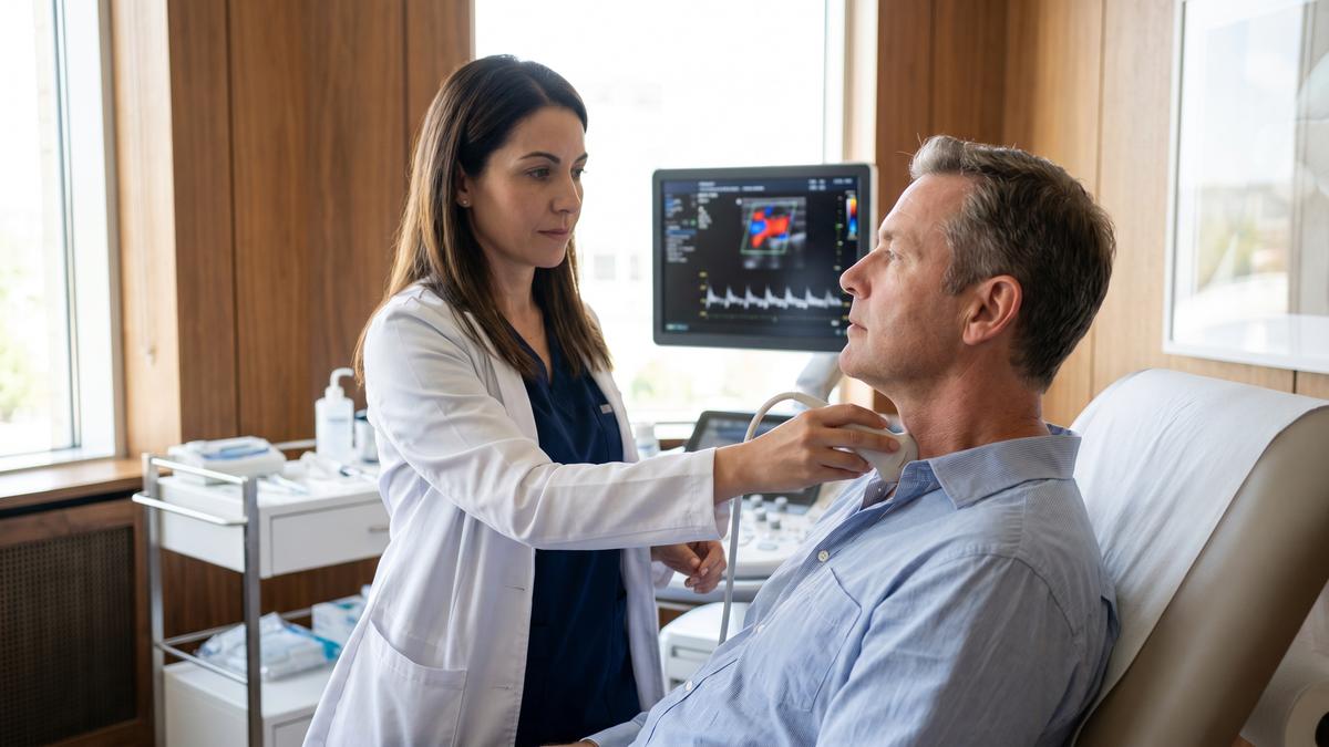
Ultrasound is a noninvasive test used to diagnose and monitor conditions affecting the blood vessels. Specifically, it's used for diagnosing aneurysms (narrowing of vessels), blockages, and blood clots.
Ultrasound technology uses sound waves to produce real-time images of arteries and veins. It then measures the speed at which these waves bounce back from tissue being examined, known as echolocation.
Aneurysms
Medical ultrasound is a noninvasive imaging procedure that utilizes high-frequency sound waves to examine the interior of the body. It can help detect conditions affecting blood vessels and other organs, such as tumors; additionally, it may identify changes in tissue appearance.
Aneurysms, which can occur anywhere on the body, are a type of vascular condition caused by a weak spot in an artery wall that grows and bulges. They may present with various symptoms but the most common one being pain at the site of the aneurysm.
Some aneurysms may not cause any symptoms at all, and in these cases there's usually no need for treatment unless they grow or rupture. Nonetheless, you should still be monitored by your physician in case these small bulges develop into larger problems.
Your healthcare provider can detect aneurysms through physical examination, x-rays and ultrasound imaging. Ultrasound imaging also assists your doctor in determining the size and location of an aneurysm.
Brain Aneurysm (Brain Bleed)
If an aneurysm in the brain ruptures, it could result in subarachnoid hemorrhage - a serious form of stroke that takes place when a blood clot blocks off blood flow to the brain.
Dr Teitelbaum notes that brain bleeds can be caused by many things, including injury, infection and genetic factors. But the most frequent reason is a congenital weak spot in the artery wall which expands over time.
Other causes may include atherosclerosis, a blood vessel disease; heart disease; high cholesterol; diabetes; and smoking. People who have a family history of aneurysms are 12 to 15 times more likely to develop one themselves, Teitelbaum notes.
Large aneurysms can lead to strokes if they occlude an artery and prevent blood flow to the brain, so it's imperative that individuals get screened for brain aneurysms as soon as possible.
To reduce the risk of aneurysms, you should manage your risk factors by abstaining from tobacco and alcohol use, maintaining a healthy weight, and managing high blood pressure.
If an aneurysm has already ruptured, neurosurgery may be necessary to clip its blood vessels. This involves finding which vessels supply the aneurysm and attaching a small metal clip on its neck which stops blood flow. Although this procedure has proven successful for some people, there are risks and potential issues to consider.
Blockages
Medical ultrasound can detect blockages and other conditions affecting blood vessels. It's also used to monitor your vascular health and perform procedures like biopsies and drainages.
Diagnostic ultrasound is a non-invasive, low energy technique that creates images of internal organs and structures within the body. It's used for diagnosing diseases such as heart disease, breast cancer, kidney stones, bladder tumors and uterine fibroids.
Ultrasound is a safe and reliable imaging technique, but it has limitations. For example, it cannot accurately image certain body parts such as the lungs or head because sound waves do not travel well through air or bone. To address these concerns, your doctor may order other tests like CT scans or X-rays instead.
Angiography is an imaging test that employs X-rays and dye (contrast) to view the inside of your blood vessels. During this procedure, a small amount of dye is injected into each vessel so that they appear clearly on the test image.
Your healthcare provider may use angiography to inspect the arteries in your arms and legs for blockages. If there are, you may experience pain or discomfort; if so, medication may be prescribed by your doctor to alleviate symptoms.
A common type of artery blockage occurs due to a buildup of fat and cholesterol inside the arteries. When these deposits build up, the arteries narrow, preventing oxygen-rich blood from reaching your brain or other parts of your body - this condition is known as atherosclerosis.
If your arteries become so narrow that they cannot carry blood to your brain, you may suffer a stroke - also known as TIA or transient ischemic attack (TIA).
Functional ultrasound is a type of imaging technology used to measure and visualize blood flow in vessels like carotid arteries or abdominal aortas. It also measures tissue stiffness throughout the body, making it possible to differentiate tumors from healthy tissues. This information can be displayed as color-coded maps, black-and-white high contrast maps or overlays on anatomical images.
Blood clots
Blood clots are solid masses of blood formed when platelets (a type of blood cell) and proteins in plasma (the liquid part of your blood) stick together. They're natural and help stop bleeding when blood vessels are cut, injured, or damaged; however they usually dissipate once the injury heals.
However, occasionally blood clots will not dissolve and can prevent blood flow to vital organs like your heart and brain. This is incredibly dangerous and requires accurate diagnosis and treatment for effective relief.
Doctors use medical ultrasound to diagnose and monitor conditions affecting the blood vessels. By using sound waves combined with a computer, this technology creates pictures of inside your body which may include heart disease or kidney stones.
If your doctor suspects you of having a blood clot, they may order tests to detect D-Dimer (an enzyme responsible for helping the body clot) and other antibodies which could prevent clotting. These can be conducted in either your doctor's office or the emergency room.
If you have a blood clot, your doctor may prescribe anticoagulant medications (also referred to as "blood thinners") in order to slow the body's ability to form new clots and keep existing ones from growing larger. They'll also advise you on ways to move around so that you're not sitting for extended periods of time or lying flat on your back.
Additionally, you'll have a series of laboratory tests to monitor the clotting status of your blood. This may include a D-Dimer test and other work to detect whether you have a genetic disorder that could cause your body to clot more frequently than usual.
In some cases, your doctor may suggest catheter-directed thrombolysis as a treatment option. In this technique, they use a thin tube with sophisticated tools at its tip to reach and remove the clot, without needing large incisions.
A common blood clot is known as deep vein thrombosis (DVT). This clot forms in a large vein such as your leg or arm and can partially or completely block blood flow through it.
Treatment
Medical ultrasound is a noninvasive, safe way to examine the structures of your body. It uses high-frequency sound waves to create images of internal organs and blood vessels, such as the heart and arteries. Ultrasound can also be used to diagnose and treat various conditions like breast lumps or vascular disease.
Medical ultrasound, when performed by a qualified professional, is an noninvasive and painless way to visualize the structures inside of your body. Compared to X-rays or CT scans, it does not use radiation which could potentially harm an unborn child.
Healthcare providers also utilize vascular ultrasound to assess the health of your arteries and veins. This test can help them detect an obstruction in an artery, such as plaque buildup from fats, cell debris and calcium that causes atherosclerosis - a major risk factor for heart disease.
If a vascular ultrasound reveals that your blood flow is irregular or slow, your provider may suggest placing a stent to narrow the artery if possible. Alternatively, they could suggest using a graft to replace the affected section of blood vessel.
Vascular ultrasound can also be used to assess the success of an angioplasty or verify the viability of vein bypass surgery. It helps your provider decide the most suitable course of action when treating varicose veins or spider veins, should they exist.
You'll likely be asked to lie on an exam table or on a transducer, which is a special padded device used for examinations. A gel is applied to the area being examined and then the transducer moves over it with images captured of your body.
Your vascular ultrasound exam may take anywhere from 30 to 60 minutes, depending on the type of testing needed. Following the scan, a specialist or sonographer will review the results with you.
When having a vascular ultrasound, you will typically be asked to lie down on an examination table and drink warm water while waiting. Afterward, an injection of medication will be given in order to relax your muscles.