
- Home
- Women
- Men
- MSK
- Cardiovascular
- Screening
- About
- Book a Scan
- Blog
A discreet, painless testicular health assessment using gold-standard Doppler imaging. Get clear answers from an experienced sonographer, with a detailed written report within 24 hours.
No preparation required · Marylebone, Central London · HCPC registered sonographers
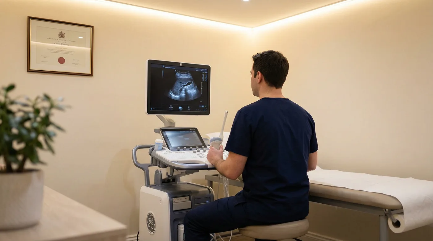
A private testicular ultrasound-scan is a non-invasive diagnostic imaging procedure that uses high-frequency sound waves to produce real-time images of the testicles, epididymis, and surrounding scrotal structures. It is the gold-standard imaging modality for scrotal pathology, recognised as such by the European Association of Urology (EAU) and the British Association of Urological Surgeons (BAUS).
The scan works by transmitting sound waves through a small handheld probe placed gently against the skin. These waves reflect off internal tissues at different rates depending on their density, and the returning echoes are converted into detailed greyscale images on screen. A colour Doppler component is added to assess blood flow within the testicular vasculature — a critical factor in distinguishing benign conditions such as epididymo-orchitis from those requiring urgent surgical attention, such as testicular torsion.
Unlike X-rays or CT scans, testicular ultrasound uses no ionising radiation, making it entirely safe and suitable for repeated use when monitoring is required. The scan offers sensitivity exceeding 95% for detecting intratesticular masses — information that physical examination alone cannot deliver.
Why book privately rather than waiting for an NHS referral?
| Feature | Detail |
|---|---|
| Scan type | Greyscale B-mode + colour Doppler |
| Duration | 15–30 minutes |
| Preparation | None required |
| Radiation | None — uses sound waves only |
| Sensitivity for masses | >95% (EAU guidelines) |
| Report | Written report within 24 hours |
| Price | £235 all-inclusive |
| GP referral | Not required |
Key statistics
Noticing a lump, experiencing discomfort, or feeling a change in your testicles can be genuinely frightening. Many men delay seeking help — embarrassment plays a part, and so does fear of what might be found. Waiting, however, does not make symptoms disappear. It prolongs anxiety and, in cases where something serious is present, it can delay treatment that makes a real difference to outcomes.
Testicular cancer is the most common cancer in men aged 15–35 in the UK, yet it is also one of the most treatable when caught early. A private testicular ultrasound-scan gives you a definitive answer — quickly, privately, and without the stress of NHS waiting lists that can stretch to six weeks or more.
When to seek emergency care
Sudden, severe testicular pain — particularly if accompanied by nausea, vomiting, or swelling — may indicate testicular torsion, a surgical emergency. Absent blood flow on Doppler is the critical finding. Go directly to A&E or call 999. Do not wait for a private appointment.
The scan investigates a wide range of scrotal and testicular conditions — most of which are benign, but all of which benefit from accurate diagnosis. Understanding what the scan can and cannot detect helps you make an informed decision about whether it is the right investigation for your symptoms.
| Condition | What the scan shows | Urgency |
|---|---|---|
| Testicular cancer | Hypoechoic (darker) intratesticular mass; vascularity on Doppler | Urgent |
| Epididymal cysts & spermatoceles | Fluid-filled sac attached to epididymis; anechoic, smooth walls | Benign |
| Varicocele | Dilated pampiniform plexus veins (>3mm); retrograde flow on Doppler Valsalva | Monitor |
| Hydrocele | Anechoic fluid surrounding the testicle within the tunica vaginalis | Benign |
| Orchitis & epididymo-orchitis | Enlarged, hypoechoic testicle; hyperaemia (increased blood flow) on Doppler | Treatment needed |
| Testicular torsion | Absent or markedly reduced intratesticular blood flow on Doppler | Surgical emergency |
| Trauma assessment | Testicular rupture, haematocele, contusion; disrupted tunica albuginea | Urgent |
| Microlithiasis | Multiple small echogenic foci (<3mm) scattered within testicular parenchyma | Surveillance |
| Undescended testicle | Localises testicle in inguinal canal or abdomen; assesses size and echogenicity | Monitor |
Ultrasound is the primary tool for detecting intratesticular masses. Malignant tumours typically appear as hypoechoic lesions within the testicular parenchyma, often with increased vascularity on Doppler. Testicular cancer is the most common cancer in men aged 15–35 in the UK. When detected at stage 1, the five-year survival rate exceeds 98% — which is why early imaging matters.
Abnormal dilation of the pampiniform venous plexus, present in approximately 15% of all men and 35–40% of men investigated for infertility. Colour Doppler ultrasound is the most accurate non-invasive method of grading varicocele severity. A grade 3 varicocele (visible and palpable at rest) carries the highest association with impaired sperm parameters and may be amenable to surgical correction.
These are fluid-filled sacs attached to the epididymis. They are extremely common, almost always benign, and typically require no treatment. Ultrasound confirms their nature and distinguishes them from solid masses — providing the reassurance that a physical examination alone cannot offer.
Inflammation of the testicle (orchitis) or combined inflammation of the testicle and epididymis (epididymo-orchitis), usually caused by bacterial or viral infection. Doppler imaging demonstrates characteristic hyperaemia (increased blood flow), confirming the diagnosis and guiding antibiotic treatment. Untreated epididymo-orchitis can lead to abscess formation or testicular atrophy.
A collection of fluid within the tunica vaginalis surrounding the testicle. Ultrasound confirms the diagnosis, assesses the volume of fluid, and checks for any underlying testicular abnormality that may have caused the hydrocele to develop — a step that physical examination cannot achieve.
Tiny calcifications within the testicular parenchyma, appearing as small bright foci on ultrasound. Testicular microlithiasis (TM) is associated with a modestly increased risk of testicular germ cell tumours. Current EAU guidance recommends annual ultrasound surveillance for men with TM who also have additional risk factors such as a personal or family history of testicular cancer, cryptorchidism, or infertility.
Not every symptom carries the same clinical weight, but all of them carry the same psychological weight — the uncertainty of not knowing. The urgency cards below reflect clinical priority, not a reason to delay. Any symptom that is new, persistent, or changing warrants investigation.
Any new lump, whether painful or painless, should be evaluated promptly. Most lumps are benign, but only imaging can confirm this. Book within 24 hours.
Sudden, severe testicular pain may indicate torsion — a surgical emergency. Go to A&E immediately. Do not wait for a private appointment.
Ongoing dull aches or discomfort lasting more than a week require investigation to identify the cause, whether orchitis, epididymitis, or a varicocele.
A noticeable increase in testicular size or general scrotal swelling can indicate fluid accumulation, inflammation, or an underlying mass.
Varicoceles are present in up to 40% of men with fertility issues. A Doppler scan can detect and grade them accurately, informing treatment decisions.
Men with elevated risk (family history, prior testicular cancer, cryptorchidism, microlithiasis) benefit from periodic ultrasound surveillance.
The cost of delaying
The most common pattern we see is men waiting weeks or months to get a symptom checked, hoping it will resolve on its own. While the majority of testicular lumps are benign, only imaging can confirm this. Testicular cancer is highly treatable when caught at stage 1, with survival rates exceeding 98%. Delaying a scan prolongs anxiety and, in cases where something serious is present, it can make treatment more complex.
One of the practical advantages of a testicular ultrasound-scan is that it requires no preparation at all. No fasting, no full bladder, no special diet. You can eat and drink normally beforehand and attend at any time of day.
What to expect on arrival
Simple, straightforward, and discreet — from booking to receiving your results.
No GP referral is required. Book directly online or call 020 3633 4902. Same-day and next-day appointments are usually available. You choose the time that works for you.
We are located at 29 Weymouth Street, Marylebone, moments from Oxford Circus and Regent’s Park tube stations. The clinic is professional, private, and designed to put you at ease from the moment you walk in.
Your sonographer will take a brief clinical history, ask about your symptoms, and explain exactly what the scan involves. This is your opportunity to ask any questions. No preparation is required on your part.
You will lie on your back in a private examination room. The sonographer applies a small amount of warm ultrasound gel to the scrotum and moves the probe gently across the skin. The procedure is completely painless. Both testicles are examined and compared, with Doppler imaging used to assess blood flow. Your sonographer will explain what they are seeing throughout.
In most cases, your sonographer will share initial observations immediately after the scan. You will not be left wondering. If anything requires urgent attention, the appropriate next steps will be discussed with you before you leave.
You will receive a comprehensive written report including measurements, annotated images, descriptions of any findings, and clear clinical recommendations. The report can be sent directly to your GP or a specialist of your choice. If we identify something that requires urgent follow-up, we have established referral pathways to leading urologists in London.
Same-day and next-day appointments available. No GP referral needed.
Book Your Scan — £235 020 3633 4902One price covers everything. There are no separate charges for the written report, annotated images, or follow-up correspondence with your GP or specialist.
Private health insurance: Sonoworld accepts AXA Health, AXA Global, Healix, and WPA. Contact your insurer before booking to obtain an authorisation number. If you are unsure whether your policy covers testicular ultrasound, call us on 020 3633 4902.
A thorough assessment of the major abdominal and pelvic organs combined with a testicular check — four scan areas in one appointment.
Learn more →Our most extensive diagnostic package, covering all major organs, vascular health, thyroid, and testicular assessment in a single appointment.
Learn more →4.9 / 5 · Based on 287 verified reviews
“I had been putting off getting a lump checked for weeks because I was nervous about the whole thing. Booked with Sonoworld and got an appointment the next day. The sonographer was professional and made me feel completely at ease. Turned out to be a benign cyst. Wish I had gone sooner instead of worrying.”
David M.
Verified patient · January 2026
“Excellent service from start to finish. My GP felt something during a routine check-up and suggested a scan. The NHS wait was six weeks. I got scanned at Sonoworld within 24 hours, received my report the next day, and everything was clear. The peace of mind was worth every penny.”
James K.
Verified patient · December 2025
“I was worried about the scan being awkward, but it honestly was not a big deal at all. The sonographer was matter-of-fact and professional. The whole thing took about 20 minutes. They identified a varicocele affecting my fertility and referred me to a urologist. Really glad I got it checked.”
Ryan T.
Verified patient · November 2025
Consultant Sonographer · Sonoworld Diagnostic Services
Daniela Stan is a consultant sonographer with over 20 years of experience in NHS and private diagnostic ultrasound. She holds a Master’s degree in Medical Ultrasound, is registered with the Health and Care Professions Council (HCPC), and is a member of the British Medical Ultrasound Society (BMUS). Her clinical practice spans the full range of diagnostic ultrasound, with particular expertise in men’s health, vascular, and musculoskeletal imaging.
This page has been written and reviewed to reflect current clinical guidelines from the European Association of Urology (EAU) and the British Association of Urological Surgeons (BAUS). Clinical information is updated regularly to maintain accuracy.
Most men find the scan far less awkward than they anticipated. You will be in a private examination room with a single sonographer. A small drape covers the surrounding area, and the probe is applied gently to the skin — there is no internal examination. Our sonographers perform these scans routinely and approach them with complete professionalism. You can request a male or female sonographer when booking.
No. The scan is entirely painless. A small amount of warm ultrasound gel is applied to the skin and the probe is moved gently across the surface. There is no pressure, no needles, and no internal examination. If you have significant tenderness from an underlying condition, the sonographer will take extra care to minimise any discomfort.
Testicular ultrasound has a sensitivity exceeding 95% for detecting intratesticular masses, making it the gold-standard imaging modality for scrotal pathology according to the EAU and BAUS. Colour Doppler imaging adds further diagnostic precision by assessing blood flow, which is critical for distinguishing orchitis from torsion and for grading varicoceles accurately.
If the scan identifies a finding that requires urgent attention, your sonographer will discuss this with you before you leave and advise on the appropriate next steps. Sonoworld has established referral pathways to leading urologists in London who can see you quickly — bypassing NHS waiting lists entirely. Your written report is formatted for immediate onward referral to a specialist or your GP.
The scan itself takes 15–30 minutes. Allow approximately 45 minutes for the full appointment, which includes the initial clinical history, the scan, and the immediate discussion of findings with your sonographer.
In most cases, your sonographer will share initial observations immediately after the scan. A comprehensive written report with annotated images is delivered within 24 hours. The report is formatted for onward referral to your GP or a specialist and can be sent directly to them on your behalf.
No GP referral is required. You can book directly online or by calling 020 3633 4902. Same-day and next-day appointments are usually available. If you have a GP referral letter, bring it along — it provides useful clinical context — but it is not a prerequisite for booking or attending.
The private testicular ultrasound-scan costs £235 all-inclusive. This covers the full bilateral testicular and epididymal assessment, colour Doppler imaging, immediate verbal feedback, and a comprehensive written report with annotated images delivered within 24 hours. There are no hidden charges.
No preparation is required. You can eat and drink normally before your appointment. No fasting, no full bladder, no special clothing. Wear comfortable, loose clothing and bring any previous testicular imaging reports if you have them. That is all that is needed.
Ultrasound can identify intratesticular masses with a sensitivity exceeding 95% and can characterise their appearance (echogenicity, vascularity, margins) to assess the likelihood of malignancy. A definitive cancer diagnosis requires histopathological confirmation following surgical removal (orchidectomy). However, ultrasound is the essential first step that determines whether surgical investigation is warranted, and it does so with a high degree of accuracy.
Testicular microlithiasis (TM) refers to small calcifications within the testicular parenchyma that appear as bright foci on ultrasound. TM is found in approximately 5% of men undergoing testicular ultrasound. Current EAU guidelines indicate that TM alone, in the absence of other risk factors, does not require routine surveillance. However, men with TM who also have a personal or family history of testicular cancer, cryptorchidism, or infertility are advised to have annual ultrasound surveillance. Your sonographer will advise you on the appropriate follow-up based on your individual findings and risk profile.
29 Weymouth Street
Marylebone
London W1G 7DB
Nearest stations: Oxford Circus, Regent’s Park, Bond Street
Get Directions| Monday – Friday | 8:00 am – 7:00 pm |
| Saturday | 9:00 am – 5:00 pm |
| Sunday | By appointment |


