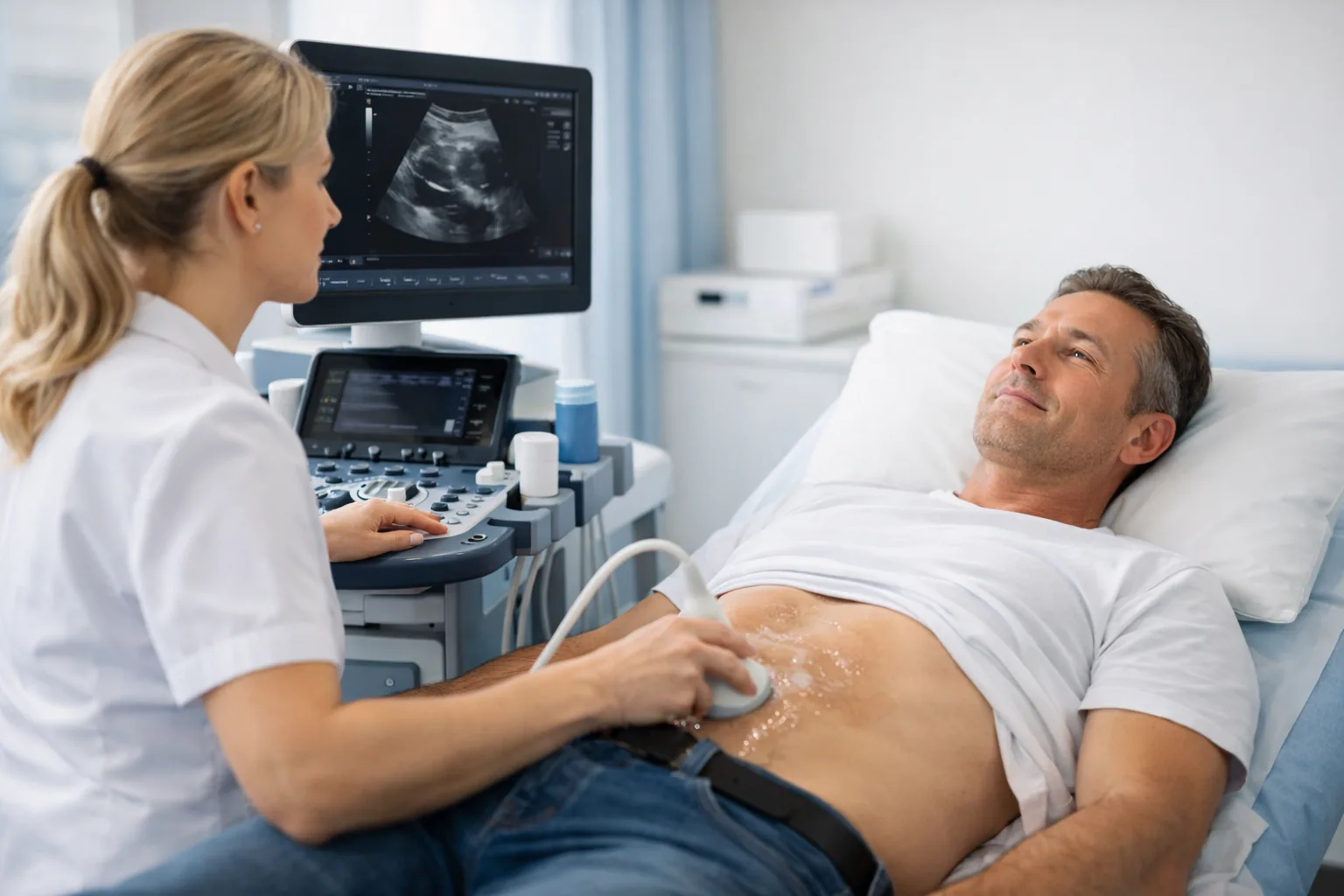
Expert vascular and cardiac ultrasound imaging at our CQC-registered clinic on Weymouth Street, in London's Harley Street Medical Area. Echocardiogram, carotid Doppler, DVT scan and aortic aneurysm screening, with same-day appointments often available and no GP referral required. Cardiovascular imaging is one part of Sonoworld's private ultrasound in London, which also covers women's health, men's health, MSK and screening scans.
CQC Registered Good No GP Referral Needed Same-Day Results Harley Street, London
Instant verbal results. Written report within 24 hours.
4Cardiovascular scan types
24hWritten report
Same DayAppointments
4.9★Doctify rating
Expert Cardiovascular Imaging
Specialist Cardiovascular Ultrasound in Central London
Cardiovascular ultrasound encompasses both cardiac imaging (echocardiography) and vascular imaging, the assessment of arteries and veins throughout the body. At Sonoworld, our specialist sonographers use high-resolution GE ultrasound equipment to provide accurate, real-time assessment of your heart and vascular system.
All cardiovascular scans are performed by HCPC-registered specialist sonographers. A comprehensive written report is delivered within 24 hours, and no GP referral is required. Same-day appointments are often available for most cardiovascular scans at our Weymouth Street clinic.
Instant verbal results and a written report within 24 hours.
No GP referral required: self-refer online or by phone.
Transparent all-inclusive pricing with no hidden fees.
Our Cardiovascular Scans
Our Cardiovascular Ultrasound Scans
Choose the scan that matches your clinical need. If you are unsure which scan is right for you, call our team for guidance.
Cardiac Ultrasound
Private Echocardiogram
A detailed ultrasound examination of the heart, assessing cardiac structure, function, valves, and chambers. The gold-standard investigation for heart murmurs, palpitations, breathlessness, and suspected cardiomyopathy.
Arterial Imaging
Carotid Doppler Scan
Detailed Doppler assessment of the carotid arteries for stenosis, plaque, and atherosclerosis. The first-line investigation for stroke risk assessment, TIA, and carotid bruit. Results available same day.
Venous Imaging
DVT Ultrasound Scan
Urgent compression ultrasound for suspected deep vein thrombosis (DVT) in the leg or arm. Same-day appointments available. A negative scan provides immediate reassurance; a positive result is reported to your GP within hours.
Aortic Screening
Aortic Aneurysm Scan
Abdominal aortic aneurysm (AAA) screening: a quick, painless ultrasound that measures the diameter of the aorta. Recommended for men over 65 and anyone with a family history of aortic aneurysm or cardiovascular risk factors.
Symptom Guide
Which Cardiovascular Scan Do You Need?
Use this guide to identify the most appropriate scan for your symptoms. If you are unsure, call our clinical team for advice.
Heart Symptoms
Palpitations, breathlessness or chest pain
These symptoms may indicate a cardiac cause. An echocardiogram assesses heart structure and function and is the first-line cardiac imaging investigation.
Stroke Risk
TIA, dizziness or carotid bruit
A transient ischaemic attack (TIA) or carotid bruit detected by your GP requires urgent carotid assessment. A carotid Doppler scan assesses stenosis and stroke risk.
Leg Swelling
Calf pain, swelling or redness
Unilateral leg swelling, calf pain, or redness after travel or surgery may indicate DVT. A DVT ultrasound scan is the gold-standard investigation and can be performed same day.
Preventive Screening
Family history or cardiovascular risk factors
Men over 65, smokers, and those with a family history of aortic aneurysm should consider an aortic aneurysm scan. Early detection is life-saving.
How It Works
What Happens at Your Cardiovascular Scan
1Book Online or by Phone
Book your scan online or call 020 3633 4902. Same-day appointments are available for most cardiovascular scans, including urgent DVT assessment.
2Arrive at Our Harley Street Clinic
Our specialist sonographer will take a brief clinical history before the scan. No fasting is required for most cardiovascular scans (echocardiogram patients should avoid a heavy meal beforehand).
3Expert Ultrasound Examination
Your scan is performed using high-resolution GE ultrasound equipment by an HCPC-registered specialist sonographer. The examination is painless and typically takes 20–45 minutes.
4Immediate Results & Written Report
Your sonographer provides immediate verbal feedback after the scan. A comprehensive written report is emailed to you within 24 hours and can be shared with your GP or cardiologist.
Patient Guides
Cardiovascular Health Resources
Education
What Is an Echocardiogram?
A comprehensive guide to echocardiography: what it shows, how it is performed, and what conditions it can diagnose.