Carotid ultrasound is a noninvasive test that utilizes sound waves to create pictures of the insides of your carotid arteries. This noninvasive procedure can help diagnose and treat carotid artery disease, an ailment commonly found among seniors.
Ultrasound testing is typically conducted in a doctor's office or hospital setting. The technician applies gel to your neck, where the carotid arteries are situated, then moves the transducer against your skin to send and receive sound waves.
Detection of plaque
Carotid ultrasound, a painless imaging test that uses high-frequency sound waves to create pictures of your carotid arteries in your neck, can detect plaque buildup. This information helps your doctor plan treatment options to remove this buildup and avoid stroke.
Cholesterol and other substances can clump together, form plaques, and thicken the walls of your carotid arteries. If left unchecked, these plaques could block blood flow to the brain and result in a stroke.
To detect carotid plaque, your doctor will order an ultrasound test. This is performed using a handheld transducer device that places gel over each carotid artery and gently presses against your neck.
The technician may utilize a device known as a computer to view the images. A radiologist will review these findings and create a report for your healthcare provider.
Carotid ultrasound can detect plaque and narrowing of the artery lumen (stenosis) within your carotid arteries, helping your doctor decide how serious the plaque is and which treatment option is most suitable.
If your carotid artery blockage is severe, your doctor may recommend surgery to clear out the plaque. This could involve a carotid endarterectomy.
Ultrasound can be used to diagnose carotid plaque, including ultrasound for duplex scanning, magnetic resonance angiography (MRA), and computed tomography angiography (CTA). CTA is the most accurate way of detecting calcifications within this plaque.
Other imaging modalities can detect plaque and features associated with stroke risk, such as surface irregularities, ulceration and inflammation. MRI is one of the best tools for detecting these features along with IPN and LRNC; while US and CTA provide more precise indications of ulceration.
Detection of stenosis
The development of various imaging modalities, from one-dimension to 2D and 3D models, has revolutionized the diagnosis of many serious and fatal diseases like cardiovascular disease (CVD). Early detection of chronic or debilitating artery disorders like carotid artery stenosis is crucial for timely intervention to ensure survival.
If you experience sudden and/or unexplained numbness or weakness in your upper limbs, it is recommended that you receive a carotid artery scan immediately. Patients with this symptom have an increased chance of suffering a stroke compared to those without.
If you experience temporary loss of vision in both eyes, or difficulty swallowing without an obvious reason, a carotid scan should also be conducted. These symptoms are commonly linked to carotid artery stenosis.
Spinal stenosis occurs when spaces within your spine narrow, compressing the spinal cord and nerves that branch off from it. This condition can occur anywhere along the spine but usually develops in either the lumbar or cervical (neck) region.
Stenosis can be caused by a variety of conditions. Common causes include arthritis, herniated discs, bone spurs, thickened ligaments and tumors.
Starting as low back pain, symptoms may worsen with standing or walking. They may also affect your neck or arms. Medication, physical therapy and spinal injections may reduce these symptoms.
People with a history of ischemic stroke should receive either carotid angiography or CT scan to identify whether they have a stenotic plaque in their artery. This will enable physicians to treat the stenosis before it leads to stroke. Furthermore, having regular vascular exams and making healthy lifestyle choices are essential.
Detection of occlusion
Occlusion detection is an integral factor in diagnosing peripheral artery disease. When an artery becomes blocked, it can lead to ischemia (decreased blood flow to the affected extremity) and necrosis (tissue death) within that extremity.
Peripheral artery disease is often accompanied by leg pain or cramping when walking or at rest (intermittent claudication). Other symptoms of peripheral artery disease include coldness, numbness and slow-healing ulcers on the limb; in severe cases patients may develop hematoma or gangrene in these regions.
Cardiologists should routinely assess ankle-brachial index (ABI) in all patients with a family history of atherosclerosis or who have been diagnosed with coronary artery disease or cerebrovascular disease. ABI helps predict heart disease risk for these individuals.
It is also essential to assess the occlusion of the carotid arteries, as a stenotic blockage in these vessels accounts for 20% of strokes among adults. A narrowing in these vessels of more than 50% can be significant and raises the risk of another stroke in those who already have one.
A common misdiagnosis when evaluating the internal carotid arteries is mistaking a collateral vessel for an occluded main artery. This often occurs when the collateral artery has become enlarged and supplies other efferent branches, appearing as though it were parallel to the main artery but actually running parallel to it - commonly mistaken for being a patent iliac artery.
Duplex ultrasound can improve the accuracy of minimal flow detection beyond severe stenosis and flow dynamics when there is severely calcified plaque present. Furthermore, using an ultrasound contrast agent may enhance diagnostic precision for technically challenging cases or those with complex peripheral vascular disease.
Detection of blood clots
Blood clots are gel-like masses of blood that can form in both veins (venous clots) and arteries (arterial clots). While some may stay put, others can move through the bloodstream, causing havoc. These clots pose a significant risk to health as they have the potential for being life-threatening.
Blood clotting can be caused by various conditions or medications, such as cancer medications and hormone replacement therapy, smoking, pregnancy and long hospital stays or surgeries. Age also plays a role in increasing the risk of getting a blood clot; additionally, trauma increases its likelihood.
Some blood clots are harmless, such as those formed after a cut or injury. While these usually dissolve quickly after being cut or injured, others don't dissolve at all and could potentially spread throughout your body if broken off and transported through the bloodstream to other organs like your lungs or brain.
A blood clot in your lung, known as a pulmonary embolism, is an extremely serious medical emergency that could result in heart attack, stroke or kidney issues and, unfortunately, death.
If a blood clot has formed in your heart or lung, your doctor may order tests to identify its source. They might also want to rule out other medical issues that could be contributing to the clot or bleeding.
Carotid Ultrasound: This noninvasive, painless screening test uses high-frequency sound waves to examine the carotid arteries on either side of your neck. The results can help your doctor determine if you have stenosis -- a narrowing of the carotid artery that increases stroke risk -- which narrows this artery.
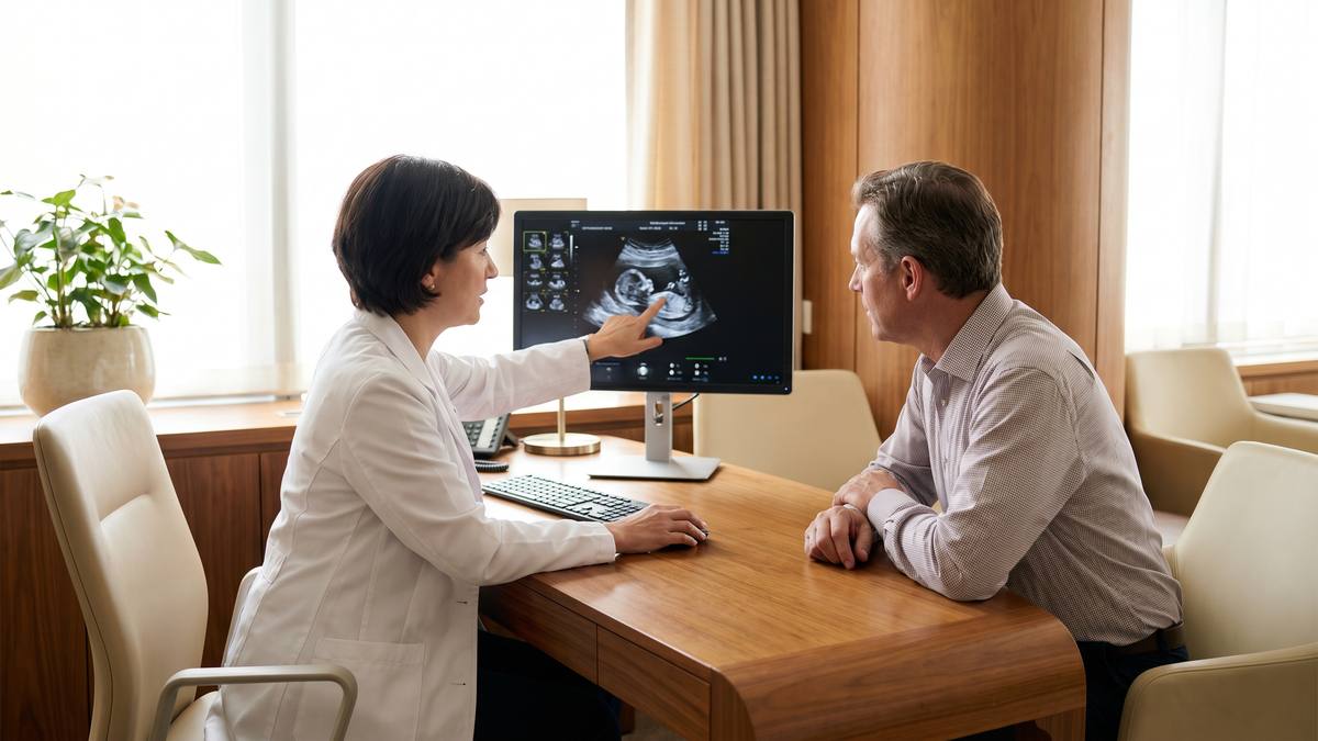
Cardiology Now offers carotid ultrasound testing, performed by a specially trained, board-certified ultrasound technologist.
Detection of vascular malformations
Carotid and peripheral vascular disease can be detected noninvasively with an ultrasound test. A technician uses a transducer, which sends ultrasonic sound waves through your skin (the same type of sound waves used by doctors to create and record ultrasound images). These waves will produce pictures of the insides of your blood vessels.
Your doctor then uses these sounds to scan a computer and assess any issues, providing them with valuable insights that may require treatments.
The most prevalent vascular malformation is a hemangioma, an abnormal growth of blood vessels. Hemangiomas typically occur in infants and usually shrink on their own over time; however, some can interfere with normal activities like vision or breathing.
Another common vascular malformation is arteriovenous malformations, or abnormal connections between arteries and veins. These can create issues with blood flow and may require lifelong medical management with specialized teams.
Vascular malformations can cause body aches, pain, swelling and issues with the blood clotting system. Furthermore, they have the potential to lead to organ damage.
When an AVM occurs in the brain, symptoms may vary. For instance, an AVM in the brain may not cause any symptoms until it bleeds, leading to headaches, seizures and muscle weakness (paralysis) on one side of the body.
A vascular malformation can be detected during pregnancy or at birth by a doctor performing an antenatal ultrasound, which detects malformations within the uterus before they are visible on standard ultrasound exams. This makes diagnosing them much simpler.