
DVT: Symptoms, Risks, and Why Early Ultrasound Matters
Medically reviewed by Daniela Stan MSc Medical Ultrasound, Consultant Sonographer & Clinical Director — July 2026
What Is DVT?
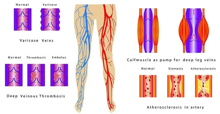
Deep vein thrombosis (DVT) is a blood clot that forms in one of the deep veins, most commonly in the leg. The clot can partially or completely block blood flow through the vein, causing pain, swelling, and redness. The most serious complication of DVT is pulmonary embolism (PE) — when part of the clot breaks off and travels to the lungs. PE is potentially fatal and requires emergency treatment.
NICE guideline NG158 estimates that DVT affects approximately 1 in 1,000 adults per year in the UK. It is more common in people over 60, but can occur at any age, particularly in the presence of risk factors.
Symptoms of DVT
DVT does not always cause symptoms — up to 50% of cases are asymptomatic. When symptoms do occur, they typically affect one leg and include:
- Pain or tenderness in the calf, thigh, or behind the knee
- Swelling of the lower leg, ankle, or foot
- Warmth and redness over the affected area
- A feeling of heaviness or aching in the leg
These symptoms overlap with other conditions — muscle strain, cellulitis, and superficial thrombophlebitis can all cause similar presentations. Ultrasound is the investigation that distinguishes between them.
Risk Factors for DVT
The main risk factors for DVT include prolonged immobility (long-haul flights, bed rest after surgery), recent surgery (particularly orthopaedic procedures), active cancer, pregnancy and the postpartum period, use of combined oral contraceptives or hormone replacement therapy, obesity, previous DVT, and inherited thrombophilia. The Wells score — a validated clinical prediction tool — combines these risk factors with clinical findings to estimate pre-test probability before imaging.
How DVT Is Diagnosed
NICE guideline NG158 recommends compression duplex ultrasound as the primary diagnostic investigation for suspected DVT. The test involves applying pressure with the ultrasound probe over the deep veins of the leg — a normal vein compresses completely, while a vein containing a clot does not. Doppler flow analysis confirms the presence or absence of blood flow. The test is non-invasive, takes approximately 20 to 30 minutes, and has a sensitivity of approximately 95% and specificity of 98% for proximal DVT.
D-dimer blood testing is used in combination with the Wells score to guide the decision to perform ultrasound — a negative D-dimer in a low-probability patient effectively rules out DVT without imaging. However, D-dimer is non-specific and is elevated in many conditions, so a positive result always requires ultrasound confirmation.
From Our Practice: The Missed DVT
A pattern we encounter is the patient who has had leg pain and swelling for several days and has been reassured by their GP that it is a muscle strain or cellulitis. They self-refer for a private DVT scan because the symptoms have not improved or have worsened. In a proportion of these cases, the scan identifies a proximal DVT — typically in the femoral or popliteal vein — that was not clinically suspected. We advise the patient to attend A&E immediately with the scan report, which supports the initiation of anticoagulation therapy. The report includes the Wells score assessment, the ultrasound findings, and a clear clinical recommendation. In these cases, the private scan has directly changed the management pathway and potentially prevented a pulmonary embolism.
Drawn from typical cases seen at our Marylebone clinic. This is a composite scenario representative of patterns we encounter regularly.
Private DVT Scanning at Sonoworld
Our DVT ultrasound scan is available from £235 at our Marylebone clinic. The scan assesses the deep veins of the leg from the groin to the calf using compression duplex ultrasound with Doppler flow analysis. Findings are explained verbally at the end of the scan and a written report is sent within 24 hours.
If the scan identifies a DVT, we will advise you to attend A&E or your GP immediately. We do not prescribe anticoagulation — that requires a clinical assessment and prescription from a doctor.
Frequently Asked Questions
How accurate is a DVT ultrasound scan?
Compression duplex ultrasound has a sensitivity of approximately 95% and specificity of 98% for proximal DVT (above the knee). Sensitivity for isolated calf DVT is lower — approximately 75% — which is why a negative scan in a high-probability patient may require repeat scanning or further investigation.
Can I have a DVT scan without a GP referral?
Yes. You can self-refer for a DVT scan at Sonoworld without a GP referral. However, if you have symptoms of pulmonary embolism — chest pain, breathlessness, or coughing up blood — you must attend A&E immediately rather than booking a scan.
How long does a DVT scan take?
The scan takes approximately 20 to 30 minutes. Results are explained verbally at the end of the appointment.
BMUS Member
ICO Registered
Ready to book your DVT ultrasound scan?
Our consultant sonographers are available Monday to Saturday at our Marylebone clinic. No GP referral needed. Instant verbal results. Written report within 24 hours.
References
- NICE. Venous thromboembolic diseases: diagnosis, management and thrombophilia testing. NG158. 2020. Available at: https://www.nice.org.uk/guidance/ng158
- Wells PS et al. Evaluation of D-dimer in the diagnosis of suspected deep-vein thrombosis. N Engl J Med. 2003;349(13):1227–1235. https://doi.org/10.1056/NEJMoa023153


