
- Home
- Women
- Men
- MSK
- Cardiovascular
- Screening
- About
- Book a Scan
- Blog
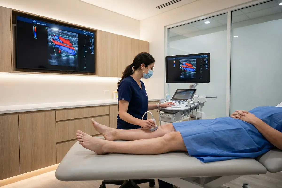
A private DVT ultrasound-scan at Sonoworld uses colour Doppler imaging to evaluate the deep veins of the leg for blood clots — with same-day results and no GP referral required.
Deep vein thrombosis (DVT) is a blood clot that forms within a deep vein — most commonly in the calf, thigh, or pelvis. Left undetected, a clot can break free and travel to the lungs, causing a pulmonary embolism, which is a life-threatening emergency.
A DVT ultrasound-scan is the first-line imaging technique recommended by NICE for suspected lower-limb DVT. Using high-frequency sound waves combined with colour Doppler technology, the examination evaluates venous compressibility and blood flow in real time — without radiation, without contrast agents, and without any preparation.
At Sonoworld, the scan is performed by HCPC-registered consultant sonographers with extensive vascular imaging experience. You receive a written report on the same day, which you can share directly with your GP or specialist.
What the DVT Ultrasound-Scan Includes
Duplex Ultrasound Accuracy
Duplex ultrasound (B-mode + Doppler) identifies proximal DVT — clots in the femoral and popliteal veins — with a sensitivity of approximately 95%. For isolated calf vein DVT, sensitivity is around 70%, as the veins become progressively smaller below the knee. Proximal clots carry the highest risk of pulmonary embolism, and these are the most reliably detected by ultrasound.
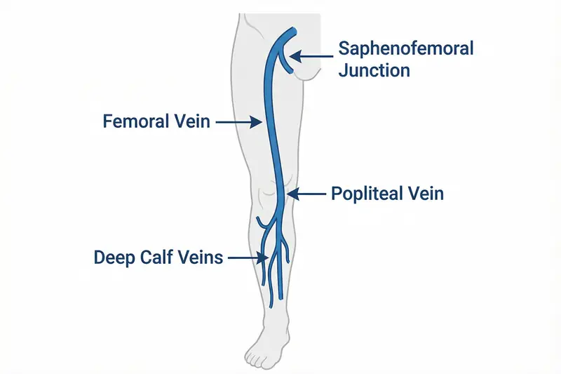
The examination follows the deep venous system from the groin to the calf, assessing each segment for compressibility, flow characteristics, and the presence of intraluminal thrombus.
| Vein / Structure | Location | Clinical Significance |
|---|---|---|
| Common Femoral Vein | Groin / upper thigh | Primary proximal DVT site; highest PE risk |
| Saphenofemoral Junction | Groin | Junction of great saphenous and femoral veins; thrombus extension point |
| Femoral Vein (mid-thigh) | Thigh | Proximal DVT; high PE risk if non-compressible |
| Popliteal Vein | Behind the knee | Proximal DVT; commonly involved in symptomatic presentations |
| Deep Calf Veins | Lower leg | Distal DVT; lower PE risk but can propagate proximally |
Colour Doppler Assessment
Colour Doppler maps the direction and velocity of blood flow within each vein. A normal vein shows spontaneous, phasic flow that augments with calf compression. Absent or reduced flow, combined with non-compressibility of the vein wall, indicates the presence of thrombus. The combination of B-mode and Doppler findings allows the sonographer to characterise whether a clot is acute or chronic.
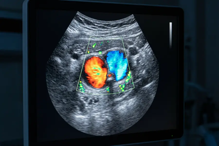
Colour Doppler image: red = arterial flow, blue = venous flow. Absence of colour in the vein indicates thrombosis.
DVT affects approximately one person in every 1,000 in the UK each year, according to NHS data. The condition becomes more common after the age of 40, though it can occur at any age. Recognising the symptoms early and understanding your personal risk profile can significantly improve outcomes.
Common Symptoms of DVT
Seek Emergency Care Immediately If You Experience
These may indicate a pulmonary embolism. Call 999 or go to A&E immediately.
Risk Factors for DVT
Related Vascular Scans
The appointment takes approximately 20 minutes for a single leg. No preparation is required — you do not need to fast or maintain a full bladder. Wear loose-fitting trousers or bring shorts, as the sonographer will need access to your leg from the groin to the ankle.
Clinical History
Your sonographer will take a brief history — duration of symptoms, relevant risk factors, and any prior DVT or anticoagulation treatment.
Positioning & Gel Application
You will lie on the examination couch with your leg exposed. A small amount of warm ultrasound gel is applied to the skin. The gel ensures full contact between the probe and the skin surface.
Compression & Doppler Imaging
The sonographer traces the deep venous system from the groin to the calf. Gentle probe pressure tests venous compressibility — a normal vein collapses fully. Colour Doppler confirms flow patterns and identifies any filling defects.
Verbal Feedback
Your sonographer will explain the findings during the examination. If a clot is identified, you will be advised on the appropriate next steps — including referral to A&E if clinically urgent.
Same-Day Written Report
A detailed written report with ultrasound images is issued on the same day. The report is formatted for GP or specialist review and can be sent electronically if required.
Ready to book your DVT ultrasound-scan?
Same-day appointments available. No GP referral required. Results on the day.
The price you see is the price you pay — no hidden extras, no referral fees. All prices include the scan, verbal feedback, written report, and ultrasound images.
Insurance & Self-Pay
Sonoworld accepts self-pay patients. If you have private health insurance, contact your insurer for pre-authorisation before booking. We can provide an invoice and report for insurance reimbursement claims.
No referral is required. You can book directly online or by telephone. If a DVT is confirmed, your sonographer will advise you on the appropriate next steps, which may include presenting to A&E or contacting your GP for anticoagulation treatment.
No preparation is required. You do not need to fast, maintain a full bladder, or avoid any medications. Wear or bring loose-fitting clothing that allows easy access to your leg from the groin to the ankle — shorts or loose trousers are ideal.
If a proximal DVT is identified, your sonographer will advise you to attend A&E or contact your GP immediately, as anticoagulation treatment is typically required. A written report will be provided for the attending clinician. For isolated calf vein DVT, management decisions are made on a clinical basis by your GP or specialist.
Yes. A bilateral DVT scan (both legs) is available for £350. This is recommended when symptoms are present in both limbs, or when clinical risk factors suggest bilateral involvement. The examination follows the same protocol for each leg.
Duplex ultrasound identifies proximal DVT (femoral and popliteal veins) with a sensitivity of approximately 95%. For isolated calf vein DVT, sensitivity is around 70%, as the veins become progressively smaller below the knee. A negative scan in a low-risk patient effectively excludes clinically significant DVT.
Your written report is issued on the same day as your appointment. The report includes the sonographer's findings, a clinical summary, and representative ultrasound images. It can be emailed directly to you or to your GP.
The scan is non-invasive and generally painless. You may feel mild pressure when the sonographer compresses the vein with the probe — this is the compression test used to assess venous compressibility. If your leg is already tender or swollen, this pressure may cause some temporary discomfort.
Sonoworld — Harley Street, London
Opening Hours
| Monday – Friday | 08:00 – 18:00 |
| Saturday | 09:00 – 16:00 |
| Sunday | Closed |
Nearest Stations


