Ultrasound is a key imaging modality in diagnosing and managing liver diseases. It is non-invasive and cost-effective, provides real-time imaging of the liver, and is therefore an important tool in the diagnosis, treatment, and follow-up of liver diseases.
In this article, we will discuss how ultrasound is used to diagnose and monitor liver disease.
Definition of ultrasound
Ultrasound is a type of imaging test used to scan the body and provide a visual representation of organs, tissues, or blood flow. It is also known as a sonogram or sonography. Ultrasound works by sending high-frequency sound waves through the body which are then reflected back and recorded. The sound waves produce images that can be used for diagnosis and monitoring.
Ultrasound can be used to diagnose a variety of conditions affecting the liver, such as inflammation, liver cysts, tumors, stones, gallstones, and masses. It can also be used to assess scarring from cirrhosis or other chronic diseases and measure the size, number, and shape of sections of the liver. Ultrasound is considered a noninvasive procedure as it does not involve radiation or incisions into the body; however it does require contact with skin through use of specialized probes attached to an ultrasound machine.
Overview of liver disease
Liver disease, also known as hepatic disease, refers to any form of damage to the liver that impairs its normal functioning. Damage can be caused by viruses and other infectious agents, medications or toxins, autoimmune diseases and genetics. It can result in poor liver function, inflammation (hepatitis), scarring of the liver tissue (cirrhosis), complete loss of liver function (liver failure) or cancer of the liver. Other forms of damage cause changes in the size and structure of the liver which may be detectable by imaging tests such as ultrasound.
Ultrasound has become an increasingly important tool in diagnosing and monitoring hepatic diseases due to its ability to produce detailed images that can detect abnormalities while providing fast results. Ultrasound provides a non-invasive method for assessing changes in size, shape and texture that are indicative of certain types of hepatic diseases. Ultrasound can be used for the diagnosis of viral infection, fatty infiltration and cirrhosis as well as monitoring changes in progression over time. It is also useful for evaluating bleeding disorders or for imaging prior to transplantation surgery or biopsy procedures.
Diagnosis
Ultrasound is a non-invasive imaging technique often used to diagnose and monitor liver diseases such as cirrhosis, fatty liver, and cysts. It uses sound waves to create detailed images of the internal structures of the liver. These images can help provide a clearer and more accurate diagnosis.
This article will focus on the diagnosis process for liver disease using ultrasound.
Ultrasound imaging of the liver
Ultrasound imaging of the liver is used to diagnose and monitor many types of liver disease, including cirrhosis, fatty liver disease, cancer, and other conditions. It can provide detailed images of the liver anatomy and any lesions or tumors that may be present.
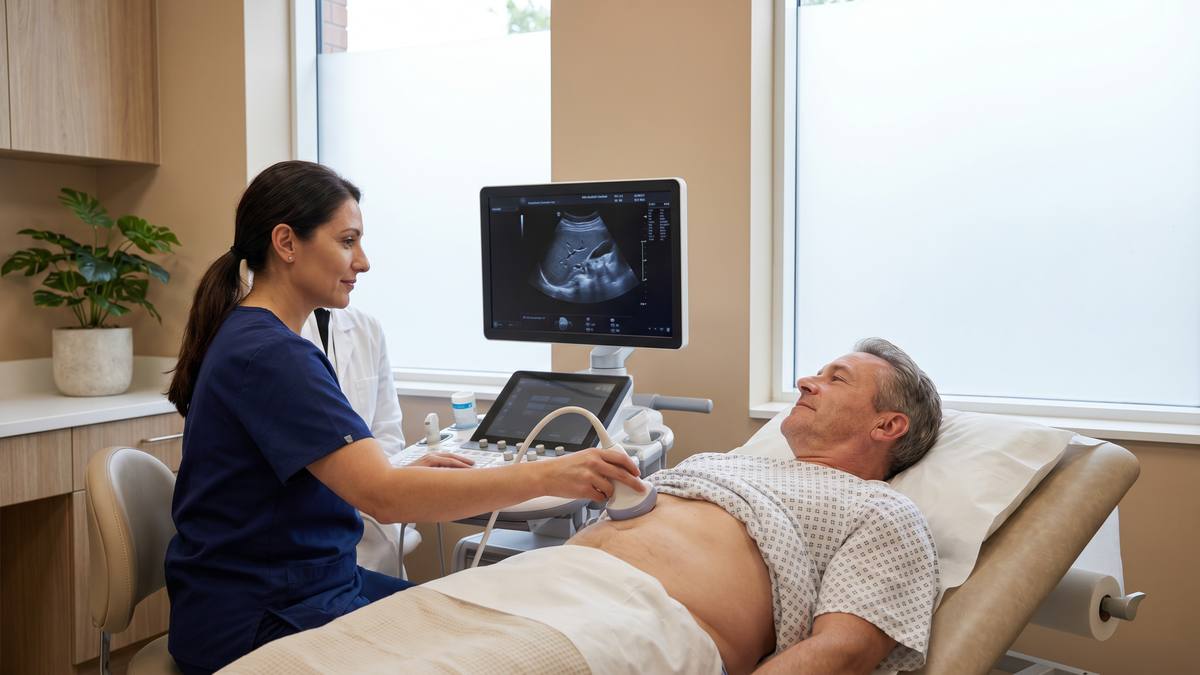
Ultrasound uses sound waves to create detailed images which can be used to examine various internal organs. The sound waves are sent into the body via an ultrasound probe which is placed on the patient's abdomen. The sound waves bounce off the internal organs within the body and are received by a transducer. These signals are then sent to a computer where they are analyzed and converted into an image for doctors to analyze and assess.
Ultrasound imaging of the liver can reveal changes in size or shape caused by inflammation or tumor growth as well as any abnormalities in blood flow inside the organ. Additionally, it can detect fluid accumulation due to portal hypertension or other causes. An ultrasound provides critical information about how a condition is affecting your liver health allowing for earlier detection and more accurate treatment decisions for medical care providers.
Evaluation of the liver using ultrasound
Ultrasound is an imaging tool used to evaluate the size, shape, structure and function of the liver. Ultrasound works by sending sound waves which are then converted into images on a computer monitor. This allows medical professionals to diagnose, monitor and treat a variety of conditions that affect the liver.
When evaluating the liver using ultrasound, several parameters are considered. These include liver size and shape, surface texture (smooth or nodular), echogenicity (bright or dull) and presence of any masses or infection in the liver. Doctors also use Doppler ultrasound to evaluate blood flow through the liver and surrounding vessels.
Abnormalities detected on a patient’s ultrasonography may be further investigated through else imaging methods such as CT scan or MRI to obtain more detailed information about specific areas of concern within the organ. Ultrasound is also used before any biopsy of tumor-like lesions in order to guide fine needles into suspicious areas for sampling tissue for study under a microscope. By combining ultrasound with other imaging tools, physicians can create comprehensive images with which they can develop accurate diagnoses and treatment plans for patients with suspected conditions affecting the liver.
Monitoring
Ultrasound has emerged as a reliable and noninvasive way for monitoring patients with chronic liver diseases like cirrhosis. It is a cost-effective and widely available imaging modality that can be used for early diagnosis and monitoring of liver diseases.
This section will provide an in-depth look at the process of monitoring liver diseases with the help of ultrasound.
Follow-up ultrasound exams
Follow-up ultrasound exams are required to carefully monitor changes in the liver. Effective follow-up ultrasound exams allow doctors to make sure treatments are working or if they need to be adjusted. Diagnostic imaging using standard ultrasounds has been shown to have promising results in monitoring the progression of liver diseases.
Standard ultrasounds provide clear images that physicians use as a reference point for future exams. Follow-up scans look for specific changes since the initial scan and allow doctors to assess how effectively treatments are working on a patient’s condition. In cases of progressive liver diseases, follow-up scans can be used to judge whether or not lifestyle changes or therapies are having any effect on developments or health of the patient’s liver.
Tests such as Advanced Liver Ultrasound, Extension Contrast Enhanced Sonography, Radionuclide Scanning, Magnetic Resonance Imaging (MRI), and Computed Tomography (CT) scans can be more detailed than typical routine ultrasound tests and provide their own particular advantages when seeking information about underlying conditions which affect the functioning of the liver. While these may provide useful additional information, often it is merely supplementing what is already captured by standard ultrasounds because of their simpler capability for routinely offering regular imaging assessments over time with a high degree of accuracy.
Use of ultrasound to monitor liver disease
Ultrasound is a useful tool to monitor a wide range of liver-related diagnostics, including visualization of the liver anatomy, examination of blood flow and assessment of disease progression. Ultrasound can also be used to detect inflammation, fibrosis or cirrhosis in the liver, as well as assessing the number and size of cystic lesions. In some cases, ultrasound has even been used to monitor the effectiveness of treatments for certain liver diseases.
Ultrasound provides detailed imagery that can help physicians diagnose complications associated with liver disease; it is generally well tolerated by patients and does not involve any exposure to radiation. During an ultrasound procedure, a machine directs high-frequency sound waves into the body and translates them into clear images on a screen. The images produced by this type of imaging can provide valuable information related to the size, shape, structure and various components within the organ being examined.
Finally, ultrasound has been found to be highly cost effective when it comes to monitoring chronic liver disease because it often removes or reduces the need for additional tests like radiography or biopsy procedures that may alternatively be required by other imaging methods.
Conclusion
In conclusion, ultrasound can be an invaluable tool for diagnosing and monitoring liver diseases in patients. As a non-invasive imaging tool, ultrasound has been proven to provide accurate and reliable results when used to diagnose and monitor liver diseases.
Furthermore, ultrasound can be used as a reliable alternative to traditional methods of liver diagnostics and monitoring. This makes it a viable option for patients who wish to avoid more invasive testing.
Benefits of ultrasound for liver disease
Ultrasound is a powerful and non-invasive imaging modality that has many advantages when used to diagnose and monitor liver disease. Ultrasound provides clinicians with real-time imaging of the liver, allowing them to monitor the health of the organ and spot any abnormalities as they occur. Ultrasound also has a distinct advantage over more invasive procedures, such as biopsy, which require you to be sedated or anesthetized before they can be performed.
The use of ultrasound in diagnosing and monitoring liver diseases has become increasingly important in recent years as it is able to accurately identify various types of diseases such as cirrhosis, fatty liver disease, infectious disorders, abdominal tumors, and cysts. As well as providing information that can help determine if a patient requires interventional therapies or other treatments for their condition.
Ultrasound is also invaluable for keeping tabs on how well existing treatments are working over time by making it easier to spot changes in the receptor tissues. This can save both time and money by identifying potential problems early on before they become more costly or serious medical complications develop. Finally, ultrasound can even provide insights into overall wellbeing by helping detect larger systemic problems caused by certain conditions when evaluating for abnormal shrinkage or expansion of the organ itself.
Limitations of ultrasound for liver disease
Ultrasound is a widely accepted imaging technique used to diagnose and monitor liver diseases. However, this modality is limited in its sensitivity for detecting smaller focal abnormalities, like early-stage cancers. As well, ultrasound may be less effective at diagnosing diffuse diseases like cirrhosis or fatty infiltration since these changes involve the whole organ rather than a specific lesion or mass.
In addition, ultrasound does not allow for tissue sample analysis (biopsy) without an invasive procedure. This means that an accurate diagnosis of intrahepatic malignancy might require additional investigations like MRI or CT scan to establish the final diagnoses based on histological studies and cell test results.
Therefore, although ultrasound can offer useful clues as to underlying pathology in select cases, it should be used in conjunction with other imaging modalities when appropriate.