By Daniela Stan MSc Medical Ultrasound, Consultant Sonographer & Clinical Director — July 2026
The Role of Ultrasound in Pancreatic Assessment
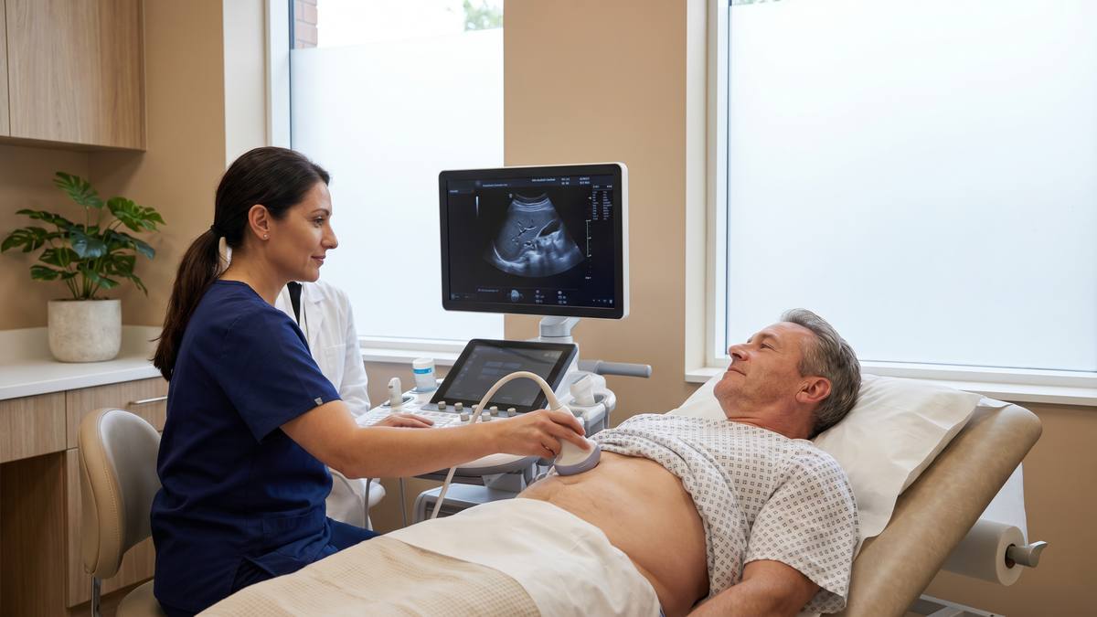
The pancreas is a retroperitoneal organ located behind the stomach, making it one of the more challenging abdominal structures to image with ultrasound. Despite this, abdominal ultrasound remains the first-line imaging investigation for suspected pancreatic pathology in many clinical scenarios, and is included as standard in our upper abdominal and full-body scan protocols. Fasting for four to six hours before the scan significantly improves pancreatic visualisation by reducing overlying bowel gas.
What Pancreatic Ultrasound Can Detect
Pancreatic ultrasound can identify dilatation of the main pancreatic duct (which may indicate obstruction by a mass or stone), pancreatic cysts (including simple cysts and mucinous cystic neoplasms), changes consistent with acute or chronic pancreatitis, and, in some cases, pancreatic masses. Secondary signs of pancreatic pathology — including biliary duct dilatation, liver metastases, and ascites — are also assessed.
Limitations of Pancreatic Ultrasound
Ultrasound has limitations in pancreatic assessment. The pancreatic tail is frequently obscured by bowel gas, even with optimal preparation. Small pancreatic masses (<2 cm) may not be visible on ultrasound. CT or MRI is typically required for definitive pancreatic assessment when ultrasound findings are inconclusive or when a pancreatic mass is suspected. Our reports clearly state when further imaging is recommended.
From Our Practice
Epigastric pain radiating to the back is one of the symptoms that patients most frequently associate with pancreatic pathology. In practice, the majority of patients with this symptom pattern have upper abdominal pathology unrelated to the pancreas — most commonly gallstones or peptic ulcer disease. Ultrasound is the appropriate first investigation in this scenario, and in most cases it identifies the cause. When the pancreas is not adequately visualised or when pancreatic pathology is suspected, we recommend CT or MRI and advise the patient to discuss urgent GP referral. Being clear about what ultrasound can and cannot show is part of our clinical responsibility.
Drawn from typical cases seen at our Marylebone clinic. This is a composite scenario representative of patterns we encounter regularly.
When Should You Have a Pancreatic Ultrasound?
A pancreatic ultrasound (as part of an upper abdominal scan) is appropriate if you have persistent epigastric or upper abdominal pain, unexplained weight loss, new-onset diabetes in a person over 50 with no family history, jaundice, or a known history of pancreatitis requiring monitoring. These symptoms require clinical assessment — see your GP if you have not already done so.
Abdominal Scanning at Sonoworld
Our upper abdominal scan (£235) includes assessment of the liver, gallbladder, bile ducts, pancreas, spleen, and kidneys. Our women's full body scan (£784) and men's full body scan (£785) include a full upper abdominal assessment as part of the seven-organ protocol.
Frequently Asked Questions
Can ultrasound detect pancreatic cancer?
Ultrasound can detect some pancreatic masses, particularly those causing biliary or pancreatic duct dilatation. However, it is not sufficiently sensitive to exclude pancreatic cancer in a symptomatic patient. If pancreatic cancer is suspected, CT or MRI is required. If your ultrasound identifies features suspicious for pancreatic pathology, the report will recommend urgent GP follow-up and further imaging.
Do I need to fast before an upper abdominal scan?
Yes. Fasting for four to six hours before the scan is essential for optimal pancreatic and gallbladder visualisation. Water is permitted. Specific preparation instructions are provided when you book.