
- Home
- Scans for Women
- Scans for Men
- Msk
- Pregnancy
- Cardiovascular
- About
- Book a Scan
- Blog
Find out exactly how your baby is positioned before labour begins. A private presentation ultrasound-scan at Sonoworld confirms whether your baby is cephalic (head-down), breech, or transverse — giving you and your midwife the information needed to plan your delivery with confidence.
Knowing your baby's position as you approach your due date matters enormously for delivery planning. A presentation ultrasound-scan confirms whether your baby is cephalic (head-down — the optimal position for vaginal birth), breech (bottom or feet first), or transverse (lying sideways across the uterus). Each position carries different implications for how your labour and delivery will be managed.
Most babies settle into a cephalic position by 36 weeks. Around 3–4% remain breech at term, and transverse lie is less common but clinically significant. Identifying a non-cephalic position early — ideally at 36–38 weeks — gives you time to discuss options with your obstetrician, consider external cephalic version (ECV), or plan a caesarean section without urgency.
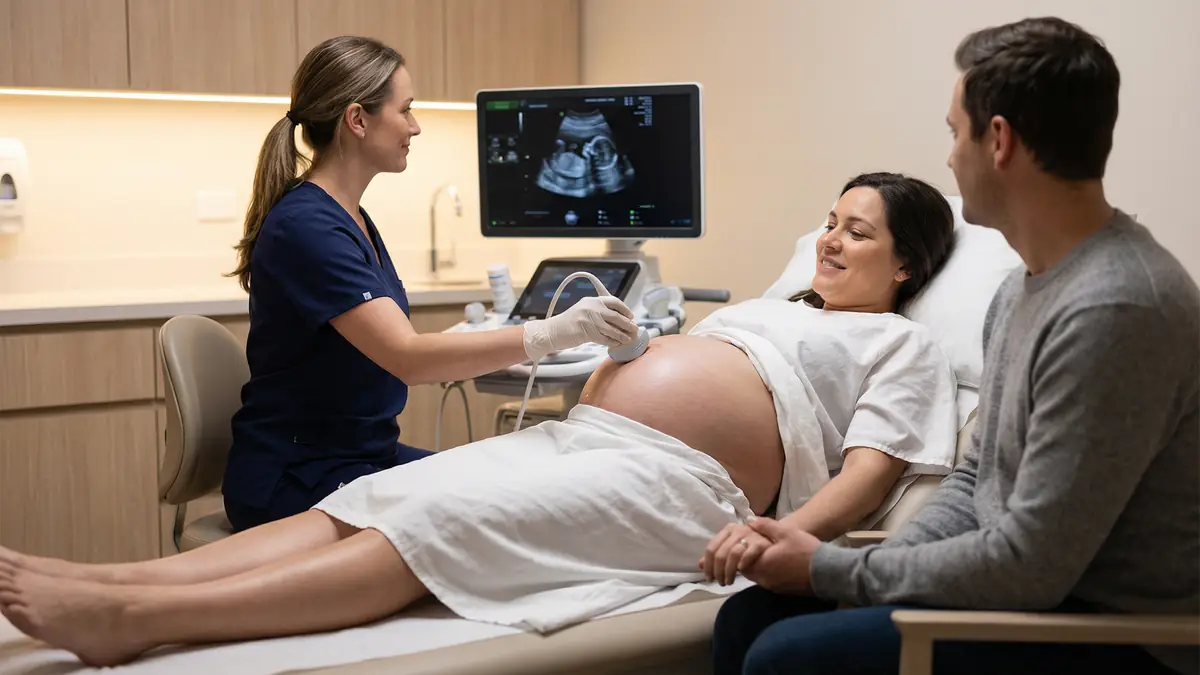
At Sonoworld, the presentation ultrasound-scan is performed by an HCPC-registered sonographer at our CQC-registered clinic at 29 Weymouth Street, Marylebone. The scan takes 15–20 minutes, requires no special preparation, and your partner is welcome to attend. You receive printed photos and a written position report to share with your midwife or obstetrician.
| Best timing | 36–38 weeks (can be performed from 32 weeks) |
| Duration | 15–20 minutes |
| Approach | Transabdominal (gel on abdomen) |
| Preparation | No special preparation required |
| Results | Immediate verbal explanation + written report |
| Photos | Printed ultrasound photos included |
| Price | £235 all-inclusive |
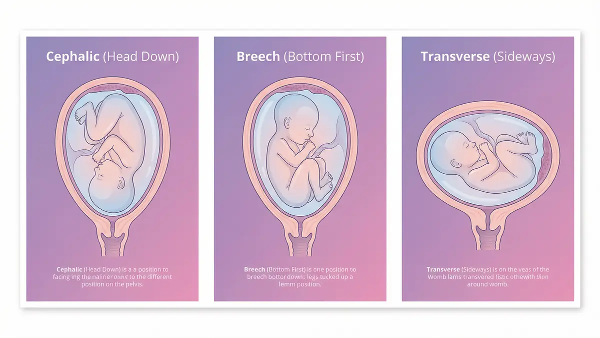
Fetal presentation refers to which part of your baby is closest to the cervix. There are three main presentations, each with distinct implications for delivery.
The head is the presenting part, positioned at the cervix. This is the optimal position for vaginal birth. Around 96–97% of babies at term are cephalic. The head can be in an anterior position (facing your spine — ideal), posterior (facing outward — can cause back labour), or transverse (sideways — usually rotates during labour).
The bottom or feet are the presenting part. Breech occurs in 3–4% of term pregnancies. There are three types: frank breech (bottom first, legs extended upward), complete breech (bottom first, legs crossed), and footling breech (one or both feet presenting). ECV can turn many breech babies at 36–37 weeks.
The baby lies horizontally across the uterus, with neither head nor bottom presenting. Transverse lie affects fewer than 1% of term pregnancies. It cannot result in a vaginal birth and requires a caesarean section if it persists at term. An oblique lie (diagonal) may also be identified and warrants close monitoring.
Babies move freely throughout pregnancy and most settle head-down between 32 and 36 weeks. After 36 weeks, the uterus becomes more crowded and spontaneous turning becomes less likely — though not impossible. A presentation scan at 36–38 weeks gives the most clinically useful information for delivery planning, while still leaving time to consider ECV if needed.
The scan covers all the key factors that influence delivery planning — not just baby's position.
| Assessment | What Is Checked | Why It Matters |
|---|---|---|
| Fetal lie | Longitudinal, transverse, or oblique | Determines whether vaginal birth is possible |
| Presentation | Cephalic, breech, or transverse | Defines the presenting part at the cervix |
| Head position | Anterior, posterior, or transverse occiput | Affects the course and duration of labour |
| Breech type | Frank, complete, or footling | Informs ECV suitability and caesarean planning |
| Placental location | Fundal, anterior, posterior, low-lying | Low-lying placenta affects delivery method |
| Amniotic fluid | AFI or deepest pool measurement | Adequate fluid is needed for safe ECV |
| Cord position | Cord location relative to cervix | Cord near cervix (vasa praevia screening) affects delivery |
| Fetal wellbeing | Fetal movement, tone, and activity | General reassurance of baby's condition |
A presentation scan is appropriate for any woman in the third trimester who wants clarity about her baby's position. These are the most common reasons patients book.
Your midwife has suggested your baby may be breech based on palpation or a previous scan. Confirm the position and explore your options — ECV, planned section, or watchful waiting.
Your baby was breech at an earlier scan and you want to know whether they have turned head-down naturally. Many babies turn spontaneously between 32 and 36 weeks.
Confirming cephalic presentation is an important step in planning a vaginal birth after caesarean. A clear position report supports your birth plan discussion with your obstetric team.
Many parents book a presentation scan at 36–38 weeks simply to confirm their baby is head-down and in a good position before labour begins. The peace of mind alone is worth it.
If your baby's movements feel different or you are unsure which way your baby is lying, a scan provides instant, definitive clarity that no amount of self-examination can match.
You simply want to know that everything is in the right position before your baby arrives. A presentation scan at 38 weeks is a popular choice for parents who want to go into labour feeling prepared.
If you experience heavy vaginal bleeding, severe abdominal pain, a sudden reduction in fetal movements, or fluid loss before 37 weeks, contact your maternity unit immediately rather than booking a private scan. A presentation scan is a planned, elective assessment — it is not a substitute for urgent obstetric assessment.
A presentation ultrasound-scan is performed abdominally — gel is applied to your tummy and the probe is moved gently across the surface. You do not need a full bladder, and there is no internal examination involved. Simply arrive at your appointment time.
Wear comfortable, loose clothing that allows easy access to your abdomen. A two-piece outfit (top and trousers or skirt) is ideal so you can simply lift your top rather than undress fully.
The optimal window is 36–38 weeks. By this stage, most babies have settled into their final position and the uterus is full enough that the baby is unlikely to turn spontaneously. Scanning at 36–38 weeks also leaves time to arrange ECV (performed at 36–37 weeks) if your baby is breech.
| Gestation | Reason for scan |
|---|---|
| 32–34 weeks | Early check if previous breech or high-risk pregnancy |
| 36–37 weeks | Optimal timing — leaves time for ECV if breech |
| 37–38 weeks | Standard presentation check before birth plan finalised |
| 38–40 weeks | Late check for peace of mind or if position uncertain |
Your sonographer will explain the type of breech position and what it means. You will receive a written report to take to your obstetric team. Options typically include external cephalic version (ECV), planned caesarean section, or in some cases a planned vaginal breech birth — your obstetrician will guide you based on your individual circumstances.
Choose a convenient appointment time online or call 020 3633 4902. Same-day and next-day appointments are usually available.
We are at 29 Weymouth Street, moments from Oxford Circus and Regent's Park stations. Your partner is very welcome. No preparation needed — just arrive.
Your sonographer will ask about your pregnancy, gestation, any previous scans, and what you would like to know. This takes just a few minutes.
Gel is applied to your abdomen and the transducer probe is moved gently across your bump. Your baby's position, placenta, and fluid are assessed in real time. The scan is comfortable and completely safe.
Your sonographer explains your baby's position on the screen, describes what it means for delivery, and answers all your questions. Nothing is left unexplained.
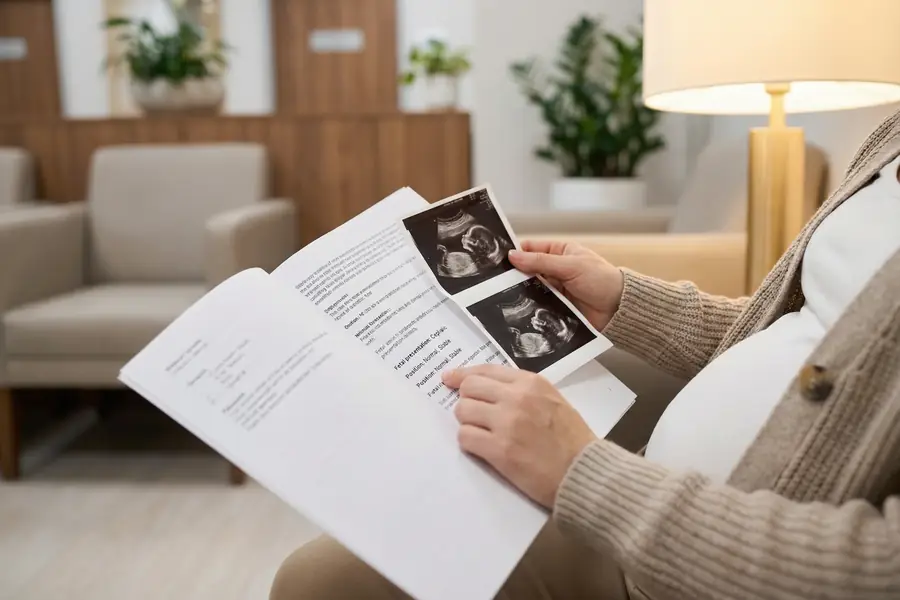
You leave with printed ultrasound photos and a detailed written position report. Share the report with your midwife or obstetrician to inform your delivery plan.
Same-day and next-day appointments are usually available. Book online in under 2 minutes or call us on 020 3633 4902.
No hidden fees. All-inclusive pricing.
One price, no hidden fees. Everything you need for confident delivery planning is included.
Many private health insurers cover presentation scans, particularly when there is a clinical indication such as suspected breech. Contact your insurer before booking to confirm coverage and obtain an authorisation number.
We accept self-pay and most major insurance providers. Call us on 020 3633 4902 to discuss your insurance options before your appointment.
020 3633 49024.9 out of 5 based on 394 verified reviews.
"Had a presentation scan at 37 weeks after my midwife thought baby might be breech. The sonographer confirmed baby was head-down in a perfect anterior position. Such a relief. The scan was quick, the report was detailed, and it really helped me feel confident going into labour."
"Baby was breech at my 32-week scan so I booked again at 36 weeks to see if she'd turned. She hadn't, but the sonographer explained my options really clearly — ECV or planned section. Really helpful for making an informed decision. Ended up having a successful ECV at 37 weeks."
"Planning a VBAC so wanted to confirm baby was head-down before labour started. Presentation scan at 38 weeks showed baby perfectly positioned and placenta well out of the way. So reassuring. Same-day appointment and lovely, supportive staff."
The ideal time is 36–38 weeks. By 36 weeks, most babies have settled into their final position and the uterus is full enough that spontaneous turning is unlikely. Scanning at this stage also leaves time to arrange ECV if your baby is breech — ECV is most effective at 36–37 weeks. If you have a high-risk pregnancy or a previous breech baby, an earlier scan at 32–34 weeks can also be useful.
Cephalic presentation means your baby's head is the presenting part — the part closest to the cervix. This is the optimal position for vaginal birth. Around 96–97% of babies at term are cephalic. Within cephalic presentation, the head can be in an anterior position (facing your spine — ideal), posterior (facing outward — can cause back labour), or transverse (sideways — usually rotates during labour).
Breech means your baby's bottom or feet are the presenting part rather than the head. It occurs in 3–4% of term pregnancies. There are three types: frank breech (bottom first, legs extended upward), complete breech (bottom first with legs crossed), and footling breech (one or both feet presenting). The type matters because it affects ECV suitability and caesarean planning.
If your baby is breech, you have several options. External cephalic version (ECV) is a procedure performed by an obstetrician at 36–37 weeks that manually turns the baby from outside the abdomen. It is successful in around 50% of cases. If ECV is not suitable or unsuccessful, a planned caesarean section is the most common recommendation. In some centres, a planned vaginal breech birth is offered to carefully selected women. Your obstetric team will guide you based on your individual circumstances. Our sonographer will explain your options at the time of the scan.
Transverse lie means your baby is lying horizontally across the uterus, with neither the head nor the bottom presenting at the cervix. It is uncommon at term (less than 1% of pregnancies) and cannot result in a vaginal birth. A caesarean section is required. Transverse lie is more common earlier in pregnancy and many babies turn to a longitudinal lie before 36 weeks. If transverse lie persists at term, your obstetric team will plan accordingly.
Yes — spontaneous turning is possible after a presentation scan, though it becomes increasingly unlikely after 36 weeks as the uterus becomes more crowded. Babies found to be cephalic at 36 weeks very rarely turn to breech. Babies found to be breech at 36 weeks have around a 7–8% chance of turning spontaneously before term. If your baby was breech at your scan and you are planning ECV, the procedure will confirm position again immediately before the attempt.
Yes. In addition to confirming fetal presentation and lie, the scan also assesses placental location (checking for low-lying placenta or placenta praevia), amniotic fluid volume (important for ECV planning), cord position (screening for cord near the cervix), and general fetal wellbeing including movement and tone. You receive a written report covering all these findings.
Ultrasound-scan is over 99% accurate for determining fetal presentation at term. This compares favourably with abdominal palpation (Leopold's manoeuvres), which has an accuracy of around 70–76% in experienced hands. Ultrasound-scan also provides additional information — placental location, fluid volume, cord position — that palpation cannot assess.
Yes. Printed ultrasound photos are included in the price. At this late stage of pregnancy, the images often show your baby's face, hands, or feet in beautiful detail. Many parents treasure these final scan photos taken before birth. You will also receive a written position report to share with your midwife or obstetrician.
The all-inclusive price is £235. This covers the 15–20 minute scan, full fetal position assessment, placenta and fluid assessment, printed photos, and a detailed written report. There are no additional fees. Many private health insurers cover presentation scans when there is a clinical indication — contact your insurer before booking to check your policy.
Same-day and next-day appointments are usually available. Book online in under 2 minutes.
Book Your Presentation Scan 020 3633 490229 Weymouth Street
Marylebone
London W1G 7DB


