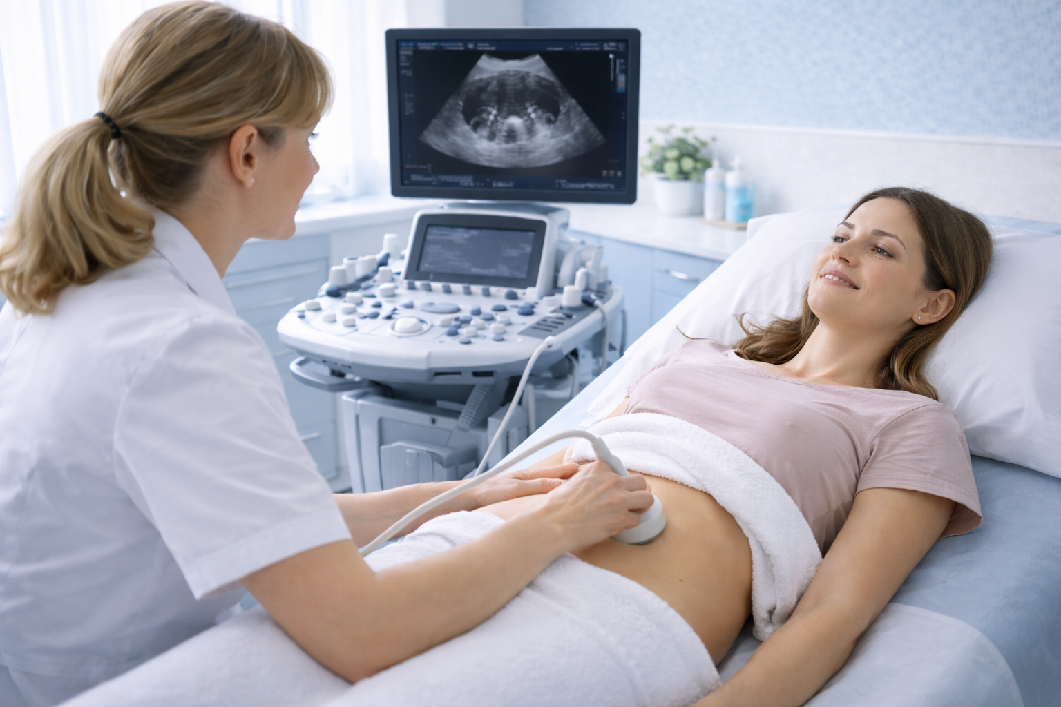
Expert transvaginal and transabdominal imaging for pelvic pain, abnormal bleeding, ovarian cysts, fibroids, PCOS, and fertility concerns. Same-day appointments at 29 Weymouth Street, Marylebone W1G.
Women's Health Scan
A pelvic ultrasound-scan is a first-line, non-invasive diagnostic imaging procedure that uses high-frequency sound waves to create real-time images of the female reproductive organs. No radiation is involved. The scan provides detailed assessment of the uterus, endometrium (womb lining), ovaries, fallopian tubes, and the surrounding pelvic structures.
Pelvic ultrasound is the imaging method of choice for investigating gynaecological symptoms. It is the recommended first-line investigation for suspected endometriosis according to NICE guideline NG73 (updated 2024), and it forms the basis of diagnosis for conditions including ovarian cysts, uterine fibroids, adenomyosis, and polycystic ovary syndrome (PCOS).
At Sonoworld, the pelvic ultrasound-scan is performed by Daniela Stan, MSc Medical Ultrasound — a consultant sonographer with over 20 years of NHS and private practice experience. You receive immediate verbal feedback during your appointment and a comprehensive written report with images within 24 hours.
Faster reassurance than typical NHS waiting times, with clear explanations throughout
Comprehensive written report and images included, suitable for GP or gynaecologist review
CQC-registered clinical environment near Harley Street (W1G) — regulated, private, and discreet
Comprehensive Assessment
The scan is tailored to your symptoms and clinical question. A comprehensive pelvic assessment covers three key areas.
The scan assesses the size, shape, and position of the uterus. The endometrial lining is measured — important for investigating heavy periods, post-menopausal bleeding, and fertility. Fibroids, polyps, and signs of adenomyosis are identified and characterised.
Conditions detectedBoth ovaries are assessed for size, follicle count, and the presence of cysts. Ovarian cysts are characterised by their appearance — simple, complex, or haemorrhagic. Signs of PCOS (polycystic ovary syndrome) and endometriomas are identified. The adnexal region is checked for masses.
Follicular trackingThe scan provides a general overview of the pelvic cavity, checking for free fluid, pelvic masses, or other incidental findings. The bladder is assessed for volume and wall appearance where clinically relevant. IUD/coil position is confirmed when requested.
Abdomen & pelvis scanDiagnostic Scope
Fluid-filled sacs on the ovaries are identified and characterised. Simple cysts, haemorrhagic cysts, dermoid cysts, and endometriomas (chocolate cysts associated with endometriosis) are assessed by size and appearance to guide management.
Fibroids (leiomyomas) are non-cancerous muscle growths in or around the uterus. Ultrasound identifies their number, size, and location — submucosal, intramural, or subserosal — which determines their impact on periods and fertility.
NICE guideline NG73 (updated November 2024) recommends transvaginal ultrasound as the first-line investigation for suspected endometriosis. The scan identifies ovarian endometriomas and signs of deep endometriosis, including involvement of the rectovaginal septum and bladder.
Ultrasound contributes to the Rotterdam criteria for PCOS diagnosis. The scan counts follicles per ovary (≥20 follicles in at least one ovary, or ovarian volume ≥10 mL) and assesses ovarian morphology alongside clinical and hormonal findings.
Adenomyosis occurs when endometrial tissue grows into the muscle wall of the uterus, causing heavy, painful periods and an enlarged uterus. Transvaginal ultrasound identifies characteristic features including myometrial cysts, asymmetric thickening, and a heterogeneous myometrium.
Endometrial polyps are small, benign growths in the womb lining that cause irregular bleeding. The scan measures endometrial thickness — a key marker in post-menopausal bleeding assessment — and identifies polyps that may require further investigation.
Procedure Types
A comprehensive pelvic ultrasound-scan typically involves both methods. Each has a specific role in building a complete diagnostic picture.
| Aspect | Transabdominal (TA) | Transvaginal (TV) |
|---|---|---|
| How it's done | Probe moved gently over the lower abdomen with gel | Small sterile probe gently inserted into the vagina |
| Best for viewing | Overall pelvic anatomy, large masses, general overview | Detailed uterus, endometrial lining, ovaries, early pregnancy |
| Preparation | Full bladder required (drink 1 litre of water 1 hour before) | Empty bladder recommended for comfort |
| Image quality | Good overview; limited detail for small structures | Superior detail — probe is closer to the organs |
| Comfort level | Fully non-invasive; very comfortable | Minimally invasive; generally comfortable; optional |
| When used | Initial assessment, large fibroids, general pelvic survey | Detailed evaluation, small cysts, endometrial assessment, PCOS |
The transvaginal scan is performed with full respect for your dignity and privacy. You are given time and privacy to undress from the waist down and are covered with a sheet throughout. The probe is approximately the width of a finger. You can decline the transvaginal component at any time — please simply let the sonographer know. If you prefer a transabdominal-only scan, this is always respected, though the level of diagnostic detail may be more limited.
When to Book
Ongoing lower abdominal or pelvic pain — whether sharp, dull, or cramping — warrants imaging to identify the cause, which may include cysts, fibroids, or endometriosis.
Heavy periods, bleeding between periods, or any post-menopausal bleeding requires investigation. Ultrasound assesses the endometrial lining and identifies structural causes.
If you are trying to conceive or planning fertility treatment, a pelvic scan provides a baseline assessment of the uterus, ovaries, and antral follicle count.
Persistent bloating, a sensation of fullness, or pressure in the lower abdomen can indicate ovarian cysts or fibroids. Ultrasound provides a clear answer quickly.
Patients who have experienced heavy, painful periods for months — sometimes years — often assume it is "just how things are". By the time they book a scan, fibroids or adenomyosis have grown significantly, narrowing the treatment options available. A pelvic ultrasound-scan takes 20–30 minutes and provides a clear answer. Early imaging means more choices.
We accept AXA, Healix, WPA, and other major insurers. Transparent pricing with no hidden fees. Self-pay patients are always welcome — the scan price of £235 includes everything.
Not sure if you need a pelvic scan or a combined scan?
If your symptoms also include upper abdominal pain, bloating after eating, or urinary symptoms, you may benefit from an Abdomen and Pelvis Ultrasound Scan instead. Call 020 3633 4902 and we will guide you.
Before Your Appointment
Preparation is straightforward. Your booking confirmation will include specific guidance, but here is what to expect.
Transparent Pricing
One price. No hidden fees. Everything you need for a complete diagnostic assessment.
Transvaginal & Transabdominal — comprehensive assessment
Insurance patients: We accept AXA, Healix, WPA, and other major insurers. Please contact us before booking to confirm your cover.
Your Appointment Journey
From booking to report — a clear, unhurried process built around your needs.
Book online and choose your preferred time slot. Same-day and next-day appointments are usually available. If you are unsure which scan to book, call 020 3633 4902 and we will guide you to the right option.
Your appointment begins with a focused conversation about your symptoms, cycle history, and any relevant medical background. This ensures the scan is targeted to your specific clinical question — not a generic protocol.
The transabdominal scan is performed first, providing an overview of the pelvic organs. The transvaginal scan follows, offering detailed assessment of the uterus, endometrium, and ovaries. The sonographer explains findings as they go.
At the end of the scan, the sonographer discusses the findings with you clearly and directly. You leave the appointment understanding what was found and what it means — not waiting anxiously for a letter.
A comprehensive written report with ultrasound images is sent to you securely within 24 hours. The report is formatted for GP and specialist review, supporting joined-up care and any referrals you may need.
Why Sonoworld
Sonoworld combines clinical expertise with a patient-centred approach, so you get the answers you need — quickly and with confidence.
Daniela Stan, MSc Medical Ultrasound, brings over 20 years of NHS and private practice experience to every scan. Her specialist knowledge in gynaecological imaging means findings are interpreted with precision and explained with clarity.
Same-day and next-day appointments are available without a GP referral. When you are worried about a symptom, waiting weeks is not an option. Sonoworld provides fast access to the imaging you need, when you need it.
Every scan includes a comprehensive written report with images, delivered within 24 hours. The report is formatted for GP and specialist review, supporting referrals, treatment planning, and informed decision-making.
Patient Reviews
"I had been suffering from severe pelvic pain for months and the NHS wait was too long. I booked with Sonoworld and was seen the same day. Daniela was incredibly gentle and explained everything during the transvaginal scan. She found an ovarian cyst and put my mind at ease straight away. The report was sent to my GP the next day."
"I booked a pelvic scan to check my coil position and investigate some irregular bleeding. The clinic is spotless and professional. Daniela was very respectful and made me feel completely at ease. She explained everything she could see on the screen. The written report was thorough and my GP was very impressed."
"I visited Sonoworld for a fertility assessment before starting IVF. The antral follicle count and ovarian assessment were done thoroughly and the results were explained in a way I could actually understand. Daniela was warm, knowledgeable, and took time to answer all my questions. Highly recommend."
FAQs
Clear answers to the questions patients ask most before their pelvic scan.
A 20-minute pelvic ultrasound-scan at Sonoworld gives you a clear answer, a detailed report, and the confidence to take the right next step — without the wait.
Sonoworld Diagnostic Services · 29 Weymouth Street, Marylebone, London W1G 7DB