
- Home
- Scans for Women
- Scans for Men
- Msk
- Pregnancy
- Cardiovascular
- About
- Book a Scan
- Blog
A detailed duplex Doppler assessment of both carotid and vertebral arteries — detecting plaque, stenosis, and measuring intima-media thickness (CIMT) to quantify your personal stroke risk. No preparation required. Written report within 24 hours.
No preparation required · No GP referral · Report within 24 hours · Not sure which scan you need?
The carotid arteries are the two large blood vessels running up either side of your neck that supply approximately 80% of the blood to your brain. When fatty deposits — called atherosclerotic plaques — build up inside their walls, the arteries narrow (stenosis) and the risk of stroke or transient ischaemic attack (TIA) increases substantially. A carotid artery duplex ultrasound-scan is the gold-standard, non-invasive method for detecting this process before it causes a life-changing event.
At Sonoworld, the scan uses high-frequency sound waves combined with colour Doppler imaging to visualise the artery walls in real time, measure blood flow velocities, characterise plaque morphology, and calculate carotid intima-media thickness (CIMT) — a validated marker of subclinical cardiovascular disease. The entire assessment covers the common carotid artery (CCA), carotid bifurcation, internal carotid artery (ICA), external carotid artery (ECA), and both vertebral arteries. No ionising radiation is used at any stage.
Unlike a standard GP appointment, which may not include vascular imaging, a private carotid scan provides a detailed, quantified picture of your arterial health that can be shared directly with your GP, cardiologist, or neurologist. The written report includes annotated images, CIMT measurements, stenosis grading using NASCET criteria, and a clinical summary — delivered within 24 hours.
| Duration | 20–30 minutes |
| Price | £235 all-inclusive |
| Preparation | None required — no fasting, no full bladder |
| Arteries assessed | Bilateral CCA, ICA, ECA, carotid bifurcation, vertebral arteries |
| Imaging modality | B-mode ultrasound + colour Doppler + spectral waveform analysis |
| Report | Verbal feedback same day; written report within 24 hours |
| GP referral | Not required |
CIMT as a stroke risk predictor
Research published in the Journal of the American Heart Association (Suita Study, 2018) found that new carotid plaque carries an adjusted hazard ratio of 2.01 for stroke. A CIMT ≥1.0 mm is associated with a 3.23-fold increase in short-term mortality following ischaemic stroke (Metabolic Brain Disease, 2021).
The carotid duplex scan at Sonoworld is a comprehensive bilateral assessment covering every clinically relevant parameter of carotid and vertebral artery health. The table below summarises what is evaluated during the examination.
| Parameter | What Is Measured | Clinical Significance |
|---|---|---|
| Carotid Intima-Media Thickness (CIMT) | Wall thickness of the common carotid artery in millimetres | Validated marker of subclinical atherosclerosis; normal <0.9 mm; ≥1.0 mm associated with 3.23× increased post-stroke mortality |
| Plaque Detection & Characterisation | Presence, size, location, echogenicity, and surface morphology | Soft/heterogeneous plaques carry higher embolic risk; ulcerated plaques associated with highest stroke risk; plaque presence has higher predictive value than CIMT alone (Vascular, 2024) |
| Stenosis Grading (NASCET) | Peak systolic velocity (PSV) and ICA/CCA ratio at bifurcation and ICA | Graded as <50%, 50–69%, 70–99%, or occlusion; ≥70% stenosis may indicate need for endarterectomy |
| Colour Doppler Flow Mapping | Direction and character of blood flow through the carotid arteries | Identifies turbulent flow, flow reversal, and areas of haemodynamic significance |
| Spectral Waveform Analysis | PSV, end-diastolic velocity (EDV), and resistance indices | Quantifies degree of stenosis; abnormal waveforms indicate proximal or distal disease |
| Vertebral Artery Assessment | Flow direction and velocity in both vertebral arteries | Detects vertebral artery stenosis and subclavian steal syndrome — a cause of posterior circulation TIA |
| Carotid Bifurcation | Morphology and flow at the CCA division point | The bifurcation is the most common site for atherosclerotic plaque formation |
The carotid duplex ultrasound-scan is the primary imaging tool for detecting and characterising the following conditions. Early identification allows for timely medical management, lifestyle intervention, or surgical referral before a stroke or TIA occurs.
Narrowing of the internal carotid artery caused by atherosclerotic plaque. Stenosis of 50–69% is associated with a 5-year stroke risk of approximately 15% in symptomatic patients; stenosis of 70–99% carries a 5-year risk of up to 26% without intervention. NICE CG68 recommends urgent carotid imaging within 24 hours of TIA or minor stroke. The duplex scan grades stenosis using NASCET criteria and identifies patients who may benefit from carotid endarterectomy or stenting.
Key finding: PSV ≥125 cm/s (ICA) suggests ≥50% stenosis; PSV ≥200 cm/s suggests ≥70% stenosis
Fatty deposits that accumulate within the arterial wall over time, driven by hypertension, hyperlipidaemia, diabetes, and smoking. The scan characterises plaque as soft (echolucent), mixed, or calcified, and assesses surface morphology for ulceration — a marker of high embolic risk. Research published in Frontiers in Cardiovascular Medicine (2023) found that plaque echogenicity and morphology are independently associated with ischaemic stroke (AUC 0.91 for risk stratification). Plaque presence carries a higher predictive value for cardiovascular events than CIMT alone (Vascular, 2024).
Key finding: Soft or heterogeneous plaques and ulcerated surfaces indicate highest stroke risk
Thickening of the carotid artery wall without visible plaque — an early, reversible stage of atherosclerosis. CIMT ≥0.9 mm is considered borderline; ≥1.0 mm is associated with significantly elevated cardiovascular risk. A 2025 study in Mymensingh Medical Journal found mean CIMT of 0.96 mm in acute ischaemic stroke patients versus 0.65 mm in controls (OR 4.53, 95% CI 1.57–13.05). CIMT measurement enables risk reclassification and can guide decisions about statin therapy or lifestyle intervention in patients with intermediate cardiovascular risk.
Key finding: CIMT ≥1.0 mm associated with 3.23-fold increased short-term mortality after stroke
Complete blockage of the internal carotid artery, typically the end-stage of progressive stenosis. Duplex ultrasound has a summary sensitivity of 0.91 (95% CI 0.81–0.97) for detecting occlusion versus digital subtraction angiography (Cochrane Review, 2022). Identification of occlusion is critical for surgical planning and for understanding the mechanism of any preceding stroke or TIA.
Key finding: Absent flow on colour Doppler with no spectral waveform confirms occlusion
Stenosis or occlusion of the vertebral arteries can cause posterior circulation TIA — presenting as vertigo, diplopia, ataxia, or drop attacks. Subclavian steal syndrome occurs when proximal subclavian artery stenosis causes retrograde flow in the ipsilateral vertebral artery, diverting blood away from the posterior brain circulation. Both conditions are identified during the vertebral artery component of the carotid duplex examination.
Key finding: Reversed or bidirectional vertebral artery flow suggests subclavian steal
A tear in the inner wall of the carotid artery that can cause a false lumen and lead to stroke, particularly in younger patients after neck trauma, chiropractic manipulation, or spontaneously. Duplex ultrasound can identify the intimal flap, double lumen, and associated haematoma, guiding urgent referral for MRI angiography and anticoagulation management.
Key finding: Intimal flap or double lumen on B-mode with abnormal Doppler waveform
A carotid artery scan is appropriate for anyone who has experienced neurological symptoms, carries known cardiovascular risk factors, or wants a proactive baseline assessment of their arterial health. The following indications are the most common reasons patients attend Sonoworld for a carotid duplex examination.
A transient ischaemic attack (TIA) or minor stroke is a medical emergency. NICE CG68 recommends carotid imaging within 24 hours of symptom onset. If you have experienced sudden weakness, speech difficulty, facial drooping, or visual disturbance — even if it resolved — seek urgent assessment.
If your GP has heard an abnormal sound (bruit) over your carotid artery with a stethoscope, this indicates turbulent blood flow and warrants urgent duplex imaging to quantify the degree of stenosis and determine whether intervention is required.
Hypertension, type 2 diabetes, hyperlipidaemia, smoking, obesity, and a family history of stroke or heart disease all accelerate carotid atherosclerosis. A baseline CIMT measurement and plaque assessment provides a quantified risk profile that can guide preventive treatment decisions.
A visible or palpable pulsation in the neck, or a mass adjacent to the carotid artery, requires imaging to exclude carotid body tumour, aneurysm, or significant atherosclerotic disease. Duplex ultrasound is the first-line investigation for these presentations.
Patients scheduled for coronary artery bypass grafting (CABG) or other major cardiac surgery often require carotid screening to identify significant stenosis that could increase perioperative stroke risk. A private carotid scan provides the necessary documentation for the surgical team.
Men and women aged 45 and over with any cardiovascular risk factor benefit from a baseline carotid scan as part of an annual health review. Serial CIMT measurements track the progression or regression of subclinical atherosclerosis in response to lifestyle changes or medication.
One of the practical advantages of a carotid artery duplex ultrasound-scan is that it requires no preparation whatsoever. You do not need to fast, drink water, or change your medications. Simply arrive at the clinic and the scan can begin immediately.
From arrival to written report, here is exactly what to expect at your Sonoworld carotid artery scan appointment.
Arrive at 29 Weymouth Street, Marylebone. You will be greeted by reception and asked to complete a short clinical history form covering your cardiovascular risk factors, current medications, and any symptoms.
Your sonographer will review your history and discuss the reason for your scan. This helps focus the examination and ensures the report addresses your specific clinical question.
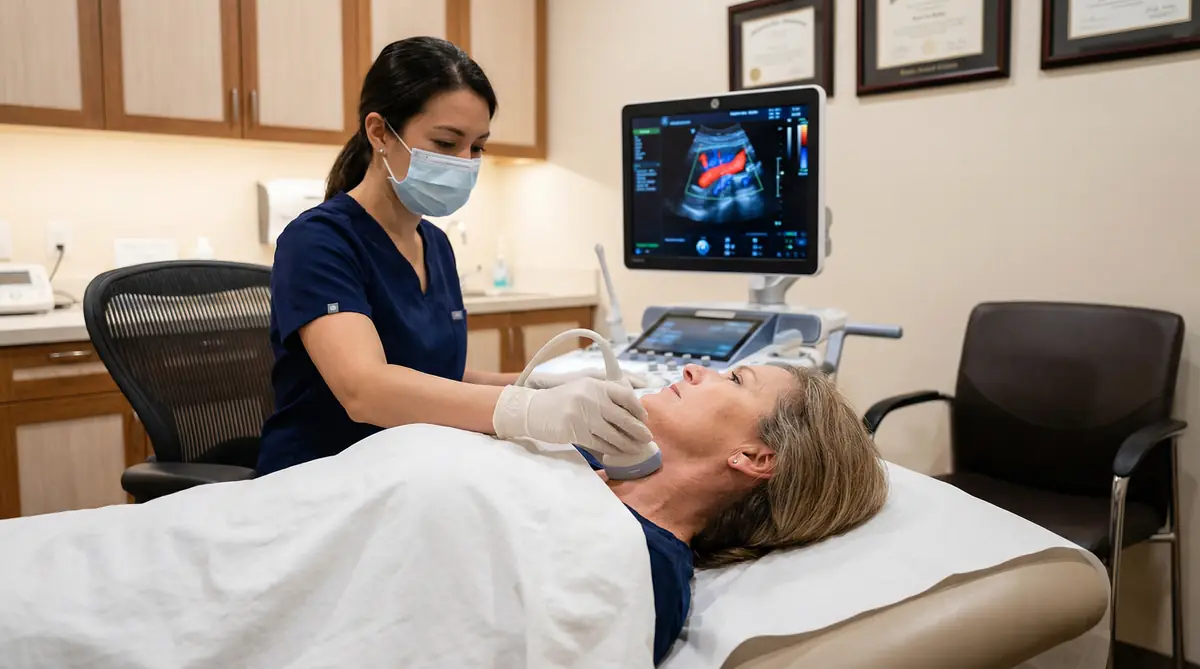
You will lie comfortably on the examination couch with your neck slightly extended. A small amount of warm ultrasound gel is applied to each side of the neck. The sonographer moves the probe gently along the carotid arteries, capturing B-mode images, colour Doppler flow maps, and spectral waveforms. Both sides are examined, followed by the vertebral arteries. The scan takes 20–30 minutes.
Carotid intima-media thickness is measured at the far wall of the common carotid artery using standardised protocol. The measurement is compared against age- and sex-specific reference ranges to provide a personalised cardiovascular risk context.
Your sonographer will explain the findings to you before you leave — what was seen, what it means clinically, and what the recommended next steps are. You will not leave the clinic without a clear understanding of your results.
A comprehensive written report with annotated images, CIMT measurements, stenosis grading, plaque characterisation, and clinical summary is delivered by email within 24 hours. The report is formatted for direct onward referral to your GP, cardiologist, or neurologist.
No. The carotid duplex scan is entirely painless. The probe is applied gently to the skin surface of the neck — there is no pressure, no needles, and no internal examination. You can resume all normal activities immediately after the scan, including driving.
Diagnostic ultrasound uses high-frequency sound waves — not ionising radiation. It is the same technology used in obstetric scanning and has an excellent safety record across decades of clinical use. There are no known risks associated with a carotid duplex examination.
Yes. You are welcome to bring a partner, family member, or friend to your appointment. They can be present in the examination room during the scan and the verbal feedback discussion.
There are no hidden charges at Sonoworld. The price below covers everything — the full bilateral carotid and vertebral artery assessment, CIMT measurement, immediate verbal feedback, and a comprehensive written report within 24 hours.
"My father had a stroke last year, so I wanted to know my own risk. The sonographer explained everything clearly — my CIMT was borderline, and the report gave my GP exactly what she needed to start me on a statin. Excellent service."
James T. — Carotid Artery Scan
"I had a TIA six months ago and my NHS follow-up was taking too long. Booked here within 24 hours, got a same-day appointment, and had a detailed written report the next morning. The scan found a 60% stenosis that my neurologist acted on immediately."
Margaret S. — Post-TIA Carotid Assessment
"Booked as part of my annual health check — I have hypertension and a family history of stroke. No plaque found and CIMT within normal range. The peace of mind alone was worth every penny. The whole process was professional and efficient."
David K. — Cardiovascular Risk Assessment
"My GP heard a bruit during a routine check and referred me. I chose to go private rather than wait. The scan was thorough, the sonographer was knowledgeable, and the report was in my inbox the following morning. Highly recommend."
Patricia W. — Carotid Bruit Investigation
Consultant Sonographer — Sonoworld Diagnostic Services, Marylebone, London
Daniela Stan is a HCPC-registered consultant sonographer with over 20 years of specialist experience in vascular and general diagnostic ultrasound. She holds a Master of Science in Medical Ultrasound and is a member of the British Medical Ultrasound Society (BMUS). Daniela has performed thousands of carotid duplex examinations and has particular expertise in CIMT measurement, plaque characterisation, and stenosis grading using NASCET criteria.
This page has been written and reviewed in accordance with NICE CG68 (Stroke and TIA), Society of Radiologists in Ultrasound (SRU) carotid duplex velocity criteria, and current peer-reviewed evidence on CIMT as a cardiovascular risk marker.
A carotid duplex ultrasound-scan detects atherosclerotic plaque, measures the degree of stenosis in the internal and common carotid arteries using NASCET criteria, and measures carotid intima-media thickness (CIMT) as a marker of subclinical cardiovascular disease. It also assesses the vertebral arteries for flow direction and obstruction.
The scan identifies soft, mixed, and calcified plaques and grades stenosis as less than 50%, 50–69%, 70–99%, or occlusion. Colour Doppler imaging visualises blood flow in real time, and spectral waveform analysis quantifies flow velocities at each arterial segment.
No. A carotid duplex ultrasound-scan is entirely painless and uses no ionising radiation. A small amount of warm gel is applied to the neck and the probe is moved gently along the carotid artery. The scan takes 20–30 minutes and you can resume all normal activities immediately afterwards, including driving.
No GP referral is required. You can book directly online or by calling 020 3633 4902. Same-day and next-day appointments are usually available. If you have a GP referral letter or previous imaging, bring it along — it provides useful clinical context — but it is not a prerequisite.
Carotid intima-media thickness (CIMT) is the combined thickness of the inner two layers of the carotid artery wall, measured in millimetres by ultrasound. A normal CIMT is below 0.9 mm; values above 1.0 mm are associated with significantly elevated cardiovascular risk.
Research published in the Journal of the American Heart Association (Suita Study, 2018) found that new carotid plaque carries an adjusted hazard ratio of 2.01 for stroke. A CIMT ≥1.0 mm is associated with a 3.23-fold increase in short-term mortality following ischaemic stroke (Metabolic Brain Disease, 2021). CIMT measurement enables risk reclassification and can guide decisions about statin therapy or lifestyle intervention in patients with intermediate cardiovascular risk.
For detecting 70–99% stenosis, carotid duplex ultrasound has a summary sensitivity of 0.85 (95% CI 0.77–0.91) and specificity of 0.98 versus digital subtraction angiography (Cochrane Review, 2022). For detecting 50–99% stenosis, sensitivity is 0.97 (95% CI 0.95–0.98).
The Society of Radiologists in Ultrasound (SRU) velocity criteria used at Sonoworld have been validated against angiography: PSV ≥125 cm/s achieves 96% sensitivity for ≥50% stenosis, and PSV ≥200 cm/s achieves 90% sensitivity and 94% specificity for ≥70% stenosis.
The private carotid artery duplex ultrasound-scan costs £235 all-inclusive. This covers bilateral carotid and vertebral artery assessment, CIMT measurement, plaque characterisation, stenosis grading, colour Doppler and spectral waveform analysis, immediate verbal feedback, and a comprehensive written report with annotated images delivered within 24 hours. There are no hidden charges.
A carotid scan assesses the arteries in the neck that supply blood to the brain, looking for plaque and stenosis that can cause stroke or TIA. An echocardiogram assesses the heart itself — its structure, valves, and pumping function. Both are complementary: carotid disease and cardiac disease often coexist, which is why Sonoworld offers a Cardiovascular Health Package combining both scans.
If the scan identifies a clinically significant finding — such as stenosis of 50% or greater, unstable plaque, or occlusion — your sonographer will discuss this with you before you leave and advise on the appropriate next steps. The written report is formatted for immediate onward referral to a vascular surgeon, neurologist, or cardiologist.
Sonoworld has established referral pathways to leading vascular specialists in London who can see you promptly. For stenosis of 70–99% in a symptomatic patient, urgent referral for consideration of carotid endarterectomy or stenting is recommended in line with NICE CG68.
Yes. Diagnostic ultrasound uses sound waves, not magnetic fields or ionising radiation, so it is completely safe for patients with pacemakers, metal implants, stents, or any other implanted devices. There are no contraindications to carotid duplex ultrasound based on implanted hardware.
A carotid duplex ultrasound-scan evaluates the extracranial vessels — it does not image the brain itself. It can identify the arterial disease that causes strokes (stenosis, plaque, occlusion) but cannot confirm brain infarction. If stroke is suspected, an MRI or CT scan of the brain is required in addition to the carotid duplex examination.
Many private health insurance policies cover diagnostic ultrasound scans. Contact your insurer before booking to confirm coverage and obtain a pre-authorisation number. Bring your policy details and authorisation number to your appointment. Sonoworld can provide a VAT receipt and clinical report in the format required by most insurers.
29 Weymouth Street
Marylebone
London W1G 7DB
Phone: 020 3633 4902
Email: info@sonoworld.co.uk
Get Directions| Monday – Friday | 08:00 – 20:00 |
| Saturday | 09:00 – 17:00 |
| Sunday | Closed |


