- Home
- Scans for Women
- Scans for Men
- Msk
- Pregnancy
- Cardiovascular
- About
- Book a Scan
- Blog
A private soft tissue ultrasound scan that identifies and characterises any lump, swelling, or mass anywhere in the body. No GP referral required. Same-day written report included.
A lumps and bumps ultrasound scan uses high-frequency sound waves to image any palpable or visible mass in the soft tissues of the body. The scan produces real-time greyscale images that reveal the size, shape, depth, internal structure, and vascularity of a lump — information that tells a consultant sonographer whether a mass is solid or fluid-filled, well-defined or irregular, and whether it contains blood flow.
Ultrasound is the first-line imaging method for superficial soft tissue masses, recommended by the British Medical Ultrasound Society (BMUS) and the Royal College of Radiologists. The vast majority of lumps are benign — lipomas, ganglion cysts, and sebaceous cysts account for the most common presentations — but characterisation matters. Knowing what a lump is removes uncertainty and guides the next step, whether that is reassurance, monitoring, or onward referral.
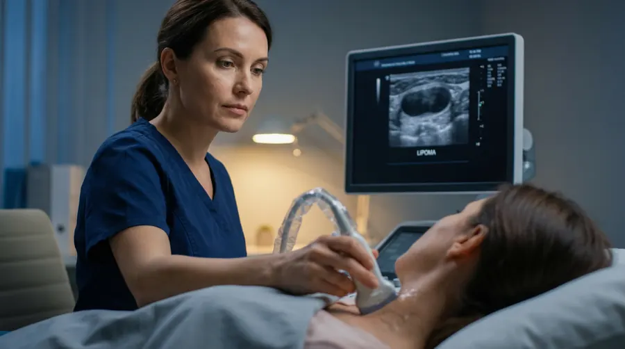
At Sonoworld, the scan is performed by Daniela Stan, a consultant sonographer with an MSc in Medical Ultrasound and over 15 years of NHS and private practice experience. The written report is issued on the same day and is accepted by GPs, surgeons, and private medical insurers.
The scan can assess lumps anywhere in the body except the breast, neck/thyroid, and joints — which have dedicated specialist scans:
Soft tissue ultrasound produces characteristic appearances for most common benign masses, allowing confident diagnosis in the majority of cases. The table below covers the most frequently encountered presentations.
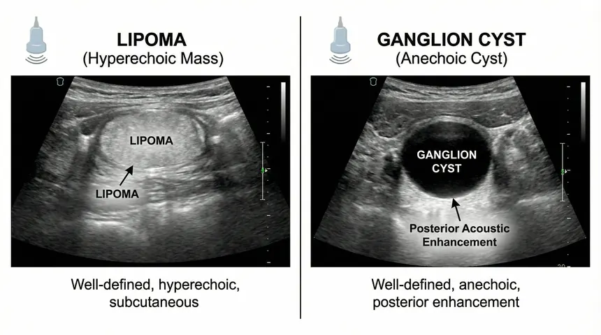
Left: lipoma (hyperechoic, well-defined) | Right: ganglion cyst (anechoic, posterior enhancement)
| Lump Type | Ultrasound Appearance | Typical Location | Clinical Significance |
|---|---|---|---|
| Lipoma | Hyperechoic or isoechoic, oval, well-defined, compressible | Subcutaneous anywhere; back, arms, thighs | Benign; no treatment required unless symptomatic |
| Ganglion cyst | Anechoic (black), round, posterior acoustic enhancement | Wrist, hand, foot, ankle | Benign; may resolve spontaneously or require aspiration |
| Sebaceous cyst | Hypoechoic, oval, superficial, may have echogenic punctum | Scalp, face, back, scrotum | Benign; excision if infected or cosmetically troublesome |
| Haematoma | Variable echogenicity depending on age; may show internal echoes | Any soft tissue after trauma | Benign; monitor for resolution; drainage if large |
| Reactive lymph node | Oval, echogenic hilum, preserved architecture, mild vascularity | Axilla, groin, neck | Usually benign; follow-up if cortex thickened or round |
| Suspicious mass | Irregular margins, heterogeneous, deep, increased vascularity | Any location, often deep | Requires urgent MRI and surgical referral |
Lumps in the breast, neck or thyroid, testicles, or within a specific joint are better assessed with dedicated specialist scans. Sonoworld offers a breast ultrasound, thyroid and neck scan, testicular scan, and a full range of MSK joint scans for these presentations.
Most people book a soft tissue scan when they notice a lump they cannot explain. Waiting weeks for an NHS referral when you have found something new and unfamiliar is a source of significant anxiety. A private scan provides answers quickly — usually within days.
The scan is appropriate for any of the following presentations:
Book as soon as possible if your lump has any of the following features:
Is your lump in a specific area? A dedicated scan may be more appropriate:
The scan takes approximately 20 minutes from start to finish. No preparation is required — you can eat, drink, and take medications as normal before attending. Wear comfortable clothing that allows easy access to the area being scanned.
The sonographer begins with a brief clinical history: when you first noticed the lump, whether it has changed, any associated symptoms such as pain, and any relevant medical or surgical history. This context directly informs the scanning protocol.
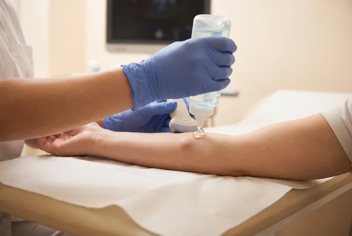
A small amount of ultrasound gel is applied to the skin over the lump. The probe is then moved across the area in multiple planes to build a complete picture of the mass. Colour Doppler is used to assess blood flow within and around the lump. The sonographer explains findings as the scan proceeds and provides immediate verbal feedback at the end.
All prices are all-inclusive. There are no hidden charges for the report, images, or verbal feedback.
Sonoworld accepts AXA, Healix, WPA, and most major private medical insurers. Please call 020 3633 4902 before booking online to ensure your insurance authorisation is processed correctly. Self-pay patients can book directly online.
Ultrasound cannot provide a tissue diagnosis — only a biopsy can confirm malignancy with certainty. However, ultrasound identifies features that are strongly associated with benign or suspicious masses. Well-defined, compressible, superficial masses with no internal vascularity are almost always benign. Irregular, deep, vascular, or rapidly growing masses require further investigation with MRI and surgical referral. The written report will state clearly whether the findings are reassuring or require urgent follow-up.
No preparation is required. Eat, drink, and take medications as normal. Wear loose clothing that allows easy access to the area being scanned. If the lump is on your torso or back, a gown will be provided.
Lumps in the breast, neck, thyroid, or testicles are better assessed with dedicated specialist scans that use specific protocols for those anatomical regions. Sonoworld offers a breast ultrasound, thyroid and neck scan, and testicular scan for these presentations. Joint lumps such as ganglion cysts at the wrist are covered by the MSK scan range. If you are unsure which scan is right for you, call 020 3633 4902 and a member of the team will advise.
A single lump scan takes approximately 20 minutes from start to finish, including the clinical history and verbal feedback. Multiple lumps will take slightly longer. The written report is issued on the same day, usually within a few hours of the appointment.
Yes. A detailed written report with annotated ultrasound images is issued on the same day. The report is produced by an HCPC-registered consultant sonographer and is accepted by GPs, surgeons, and private medical insurers. It includes the lump's measurements, ultrasound characteristics, Doppler findings, and a clinical impression with recommended next steps.
If the scan identifies features that require further investigation, the written report will include a clear recommendation — typically urgent MRI imaging and referral to a soft tissue sarcoma specialist via the two-week wait pathway. The sonographer will explain this verbally at the end of the scan and the report will provide the documentation your GP needs to make an urgent referral.
Sonoworld is located just off Harley Street in Marylebone, central London. Easily accessible from Regent's Park, Great Portland Street, and Oxford Circus Underground stations.
29 Weymouth Street
Marylebone, London W1G 7DB
Nearest stations: Regent's Park · Great Portland Street · Oxford Circus