Holter Monitor in London (Ambulatory ECG Monitoring)
If your symptoms come and go — palpitations, skipped beats, chest fluttering, dizziness, or “it happens at night / after coffee / during stress” —
a Holter monitor is designed for exactly that problem: capturing the rhythm when real life happens.
If symptoms are severe (collapse, severe chest pain, severe breathlessness at rest, stroke symptoms), treat this as urgent and seek emergency care first.
When a Holter monitor is the right choice
A Holter monitor is ideal when the main problem is timing: your symptoms don’t reliably happen during a short clinic ECG.
Holter monitoring increases the chance of capturing an episode, which is what turns worry into a specific plan.
Intermittent symptoms
Palpitations that come and go
If episodes happen in the evening, during stress, after caffeine, or randomly, a longer rhythm recording is usually more useful than repeating short ECGs.
Dizziness / near-fainting
When you need rhythm context across the day
A Holter can show if symptoms line up with rhythm changes (or if the rhythm stays normal during symptoms).
Reassurance
If anxiety is driven by “what if?”
Capturing your rhythm in normal life can be more reassuring than a single normal clinic trace — because it answers the real question: what happens outside the clinic?
After ECG
When an ECG was normal but symptoms persist
A normal ECG can be accurate — it may just mean the rhythm was normal at that moment. Holter monitoring is the typical next step for intermittent episodes.
What Holter monitoring shows (and what it doesn’t)
Holter monitoring records the heart’s electrical activity continuously. The value is the timeline:
what happens at the exact time you feel the symptom.
Holter can help identify
Arrhythmias that are intermittent (episodes you can’t catch in clinic).
Frequency of ectopic beats (how often “skipped beats” are occurring).
Correlation between symptoms and rhythm (symptom diary time stamps).
Day/night patterns including sleep-related rhythm behaviours.
Holter doesn’t image the heart
If your worry is valves, pumping function, or a murmur, Holter cannot answer that.
That’s where an ultrasound echo fits:
What is an ultrasound echo?
How the Holter appointment works
The aim is to make recording simple and low-stress: fit the monitor, explain what to do, then let you live normally while it records.
Steps (what to expect)
Step
What happens
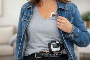
1) Fit
Electrodes are placed on the chest and connected to a small recorder. We check signal quality before you leave.
2) Record
You continue your day. We’ll explain practical points (clothing, showering, exercise guidance) for your recording period.
3) Diary
You note symptoms with approximate times (e.g., “fluttering at 14:20”). This is what allows correlation analysis.
4) Return
You return the device. We process the recording and interpret findings in context of your symptom diary.
Preparation tip: avoid heavy moisturisers on the chest on the day (they reduce electrode adhesion).
If you have previous ECGs or referral letters, bring them.
How long will I wear it?
The best recording duration depends on how often symptoms occur. If symptoms happen daily, shorter monitoring can be enough.
If they happen weekly or less, longer monitoring is often more efficient. If you want a quick decision first, compare:
Echo vs ECG
(and then choose ECG vs Holter based on symptom frequency).
Choose the right heart test (Holter vs ECG vs Echo)
Patients usually want one thing: the shortest path to certainty. Use this decision block to avoid booking the wrong test first.
Choose ECG
If you need a rhythm snapshot
Best when symptoms are happening now or you want a baseline rhythm check. ECG page:
ECG test.
Choose Holter
If symptoms are intermittent
Best when episodes come and go. Holter is designed to catch “real life” rhythms and link them to symptoms via timing.
Choose Echo
If the question is structure/pumping
Murmur, suspected valve disease, breathlessness with a structural concern, or “how well is my heart pumping?”
Start here:
What is an ultrasound echo?
“It comes and goes” → Holter. “It’s happening now” → ECG. “Is the heart pumping/valves OK?” → Echo.
Holter monitor FAQs
Short, practical answers to the questions people ask right before booking.
Is Holter monitoring painful?
No. The recorder is external and electrodes are stickers on the skin. Some people notice mild skin irritation from adhesives, especially with sensitive skin.
What should I do if I feel symptoms while wearing it?
Note the time and what you felt (e.g., “fluttering”, “dizzy”, “tight chest”) in your symptom diary. The timing is what allows correlation with the recorded rhythm.
Do I still need an ECG?
Sometimes. ECG is a snapshot and can be useful as a baseline. If symptoms are intermittent, Holter is often more informative.
For the decision logic, use: Heart monitoring hub.
When should I consider an ultrasound echo as well?
If breathlessness, a new murmur, reduced exercise tolerance, ankle swelling, or concerns about pumping/valves are central, echo may be needed.
Compare: Echo vs ECG.