Ejection Fraction & Heart Failure: What Your Numbers Mean
Left ventricular ejection fraction (LVEF) is the most important measurement used to diagnose and classify heart failure. This guide explains what your ejection fraction percentage means, how it is measured using an echocardiogram, and how it determines your treatment options.
Receiving a heart failure diagnosis or being told your heart is not pumping effectively can be an overwhelming and frightening experience. The terminology used by cardiologists can often feel confusing, leaving patients anxious about what their numbers actually mean for their daily life and future.
At Sonoworld Diagnostic Services in London, our consultant sonographers bring over 15 years of NHS and private clinical experience to every assessment. We provide rapid, same-day echocardiogram appointments without the need for a GP referral, ensuring you receive accurate diagnostic answers and a clear explanation of your results immediately. Our care is strictly aligned with the latest NICE and European Society of Cardiology guidelines.
CQC Registered
No GP Referral Needed
Same-Day Appointments
Secure online booking. Results available within 24 hours.
From £290Transparent pricing
No Radiation100% safe ultrasound
What is Ejection Fraction?
Ejection fraction (EF) is a measurement, expressed as a percentage, of how much blood the left ventricle pumps out with each contraction. The left ventricle is the heart's main pumping chamber, responsible for supplying oxygen-rich blood to the rest of the body.
Understanding the Percentage
A normal heart does not pump out 100% of the blood it holds. A healthy ejection fraction is typically between 50% and 70%. This means that with each heartbeat, the left ventricle pumps out 50% to 70% of the blood it contains. When the ejection fraction drops below 50%, it indicates that the heart muscle has become weakened or damaged, a condition known clinically as systolic dysfunction (impaired pumping ability) [1].
Why is LVEF Important?
Left ventricular ejection fraction (LVEF) is the primary parameter used by cardiologists worldwide to diagnose heart failure, determine the specific subtype of the disease, and guide treatment decisions. It is the most critical number on your echocardiogram report [2].
Heart Failure Classification by Ejection Fraction
The Universal Definition of Heart Failure classifies the condition into distinct categories based entirely on your ejection fraction percentage. This classification is crucial because the medications and treatments prescribed differ significantly between the groups [3].
Classification
Ejection Fraction (LVEF)
What it Means
Primary Treatment Focus
HFrEF (Reduced EF)
40% or less
The heart muscle is significantly weakened and cannot contract forcefully enough to pump sufficient blood. Often caused by a previous heart attack or dilated cardiomyopathy.
Guideline-directed medical therapy (GDMT) including Beta-blockers, ARNIs, MRAs, and SGLT2 inhibitors to improve survival and heart function [1].
HFmrEF (Mildly Reduced EF)
41% to 49%
A transitional or "grey zone" where the heart's pumping ability is mildly impaired. Patients in this category often share clinical characteristics with HFrEF patients [4].
Similar medications to HFrEF, focusing on preventing further decline in heart function and managing underlying conditions like coronary artery disease.
HFpEF (Preserved EF)
50% or higher
The heart pumps normally, but the muscle has become stiff and cannot relax properly to fill with enough blood between beats (diastolic dysfunction).
Managing symptoms with diuretics, controlling blood pressure, and using SGLT2 inhibitors, which have recently shown significant benefit for this group [2].
HFimpEF (Improved EF)
>40% (previously ≤40%)
A new classification for patients whose ejection fraction was previously reduced but has improved by at least 10 points due to successful medical treatment [3].
Continuing the current medical therapy to maintain the improvement and prevent relapse.
Clinical Breakthrough: SGLT2 Inhibitors
Historically, patients with Heart Failure with Preserved Ejection Fraction (HFpEF) had very few effective treatment options. However, recent landmark clinical trials have demonstrated that a class of medications called SGLT2 inhibitors (such as dapagliflozin and empagliflozin) significantly reduce the risk of cardiovascular death and hospitalisation across the entire spectrum of ejection fractions, including those above 50% [2].
Symptoms of an Abnormal Ejection Fraction
Whether your ejection fraction is reduced (HFrEF) or preserved (HFpEF), the resulting heart failure syndrome produces similar symptoms because the body is not receiving the oxygen-rich blood it requires.
When to Seek Urgent Help
If you experience sudden, severe breathlessness at rest, breathlessness accompanied by chest pain, or you cough up pink, frothy sputum, you must call 999 or attend A&E immediately. These are signs of acute pulmonary oedema (fluid in the lungs) and require emergency medical intervention.
Most Common
Breathlessness (Dyspnoea)
Feeling short of breath during everyday activities, such as walking up stairs or carrying groceries. In more advanced stages, breathlessness may occur while resting or lying flat in bed (orthopnoea).
When the heart pumps less effectively, fluid backs up in the veins, causing swelling in the ankles, lower legs, and abdomen. You may also notice sudden, unexplained weight gain.
A persistent, overwhelming sense of tiredness and weakness that does not improve with rest. This occurs because the body diverts blood away from less vital organs (like muscles) to supply the brain and heart.
Rhythm
Palpitations
A sensation that your heart is racing, fluttering, or skipping beats. A reduced ejection fraction increases the risk of developing arrhythmias, such as atrial fibrillation.
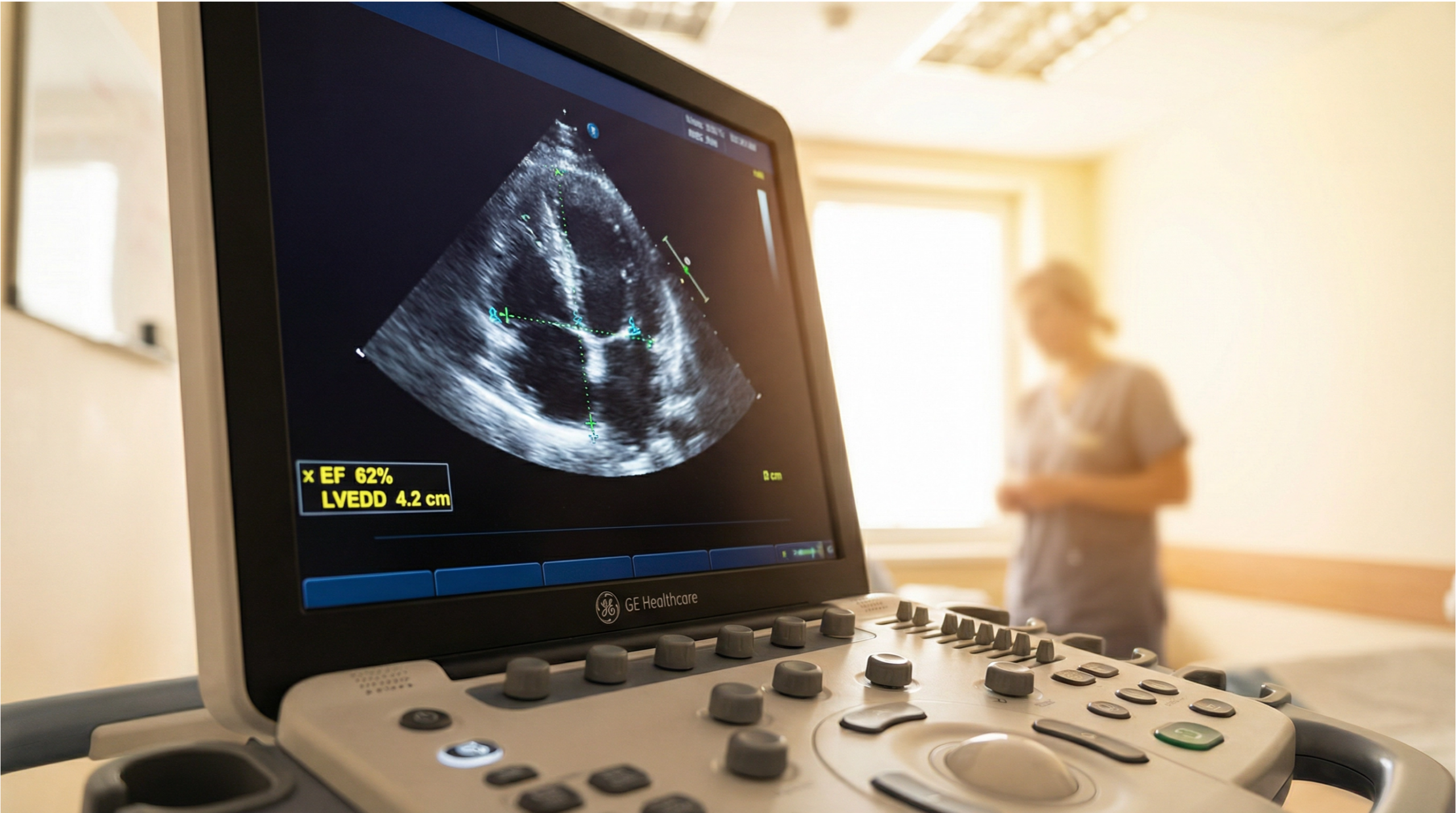
An echocardiogram (ultrasound-scan of the heart) is the gold standard, non-invasive test used to measure your left ventricular ejection fraction and diagnose heart failure. It is painless, involves no radiation, and provides immediate results.
What to Expect at Your Appointment
At Sonoworld, your assessment begins with a detailed clinical history. Our consultant sonographer will ask about your symptoms, exercise tolerance, and family history. During the ultrasound-scan, you will lie on your left side while a small probe (transducer) is moved across your chest.
Volume Measurement: The sonographer measures the volume of blood in the left ventricle when it is fully relaxed (end-diastolic volume) and when it is fully contracted (end-systolic volume).
Diastolic Function: We assess how well the heart muscle relaxes, which is essential for diagnosing HFpEF.
Valve Assessment: We check for conditions like aortic stenosis or mitral regurgitation that can cause heart failure.
A heart failure diagnosis requires a comprehensive, individualised approach. At Sonoworld, we believe in empowering patients with clear information so they can actively participate in their care plan.
Measurable Patient Goals
Treatment is not just about improving the ejection fraction number; it is about improving your quality of life. Care plans are built around measurable targets, such as:
Reducing breathlessness to allow a return to daily activities
Eliminating fluid retention and ankle swelling
Promoting reverse remodelling (the recovery of heart muscle function through medication)
Our Integrative Care Model
We provide a multidisciplinary approach to diagnostics. Our consultant sonographers work closely with referring GPs and consultant cardiologists to ensure seamless continuity of care. We adhere strictly to clinical governance standards, ensuring that your diagnostic report is comprehensive, accurate, and immediately available to guide your prescribing clinician.
Frequently Asked Questions
Can a reduced ejection fraction improve?
Yes, a reduced ejection fraction can improve significantly with the right medical treatment. The introduction of guideline-directed medical therapies, particularly the combination of beta-blockers, ARNIs, MRAs, and SGLT2 inhibitors, has been shown to promote "reverse remodelling" of the heart, leading to an increase in LVEF. Patients who experience a 10-point or greater improvement are now classified as having Heart Failure with Improved Ejection Fraction (HFimpEF).
Is an ejection fraction of 55% normal?
Yes, an ejection fraction of 55% is considered normal and healthy. A normal LVEF ranges from 50% to 70%. However, it is important to note that you can still have heart failure symptoms even with a normal ejection fraction, a condition known as Heart Failure with Preserved Ejection Fraction (HFpEF), which occurs when the heart muscle becomes stiff and cannot relax properly.
What is the difference between HFrEF and HFpEF?
HFrEF (Heart Failure with Reduced Ejection Fraction) occurs when the heart muscle is weak and cannot squeeze forcefully enough to pump blood out, resulting in an LVEF of 40% or less. HFpEF (Heart Failure with Preserved Ejection Fraction) occurs when the heart pumps normally (LVEF of 50% or higher), but the muscle is stiff and cannot relax to fill with enough blood. Both conditions cause similar symptoms but require different treatment approaches.
How often should I have my ejection fraction checked?
If you have been diagnosed with heart failure, clinical guidelines recommend regular monitoring with an echocardiogram. You should typically have a follow-up ultrasound-scan 3 to 6 months after starting or changing your heart failure medications to assess if your ejection fraction has improved. Once your condition is stable, an annual echocardiogram is usually recommended, or sooner if your symptoms worsen.
Need to check your ejection fraction?
Book a private echocardiogram at our London clinic today. No GP referral required. Fast, accurate results reported by expert consultant sonographers.
Lam, C., et al. (2021). "Classification of Heart Failure According to Ejection Fraction: JACC Review Topic of the Week." Journal of the American College of Cardiology. View source
Bozkurt, B., et al. (2021). "Universal Definition and Classification of Heart Failure." Journal of Cardiac Failure. View source
Bozkurt, B., et al. (2021). "Universal definition and classification of heart failure: a report of the Heart Failure Society of America, Heart Failure Association of the European Society of Cardiology, Japanese Heart Failure Society and Writing Committee of the Universal Definition of Heart Failure." European Journal of Heart Failure. View source
Savarese, G., et al. (2021). "Heart failure with mid-range or mildly reduced ejection fraction." Nature Reviews Cardiology. View source