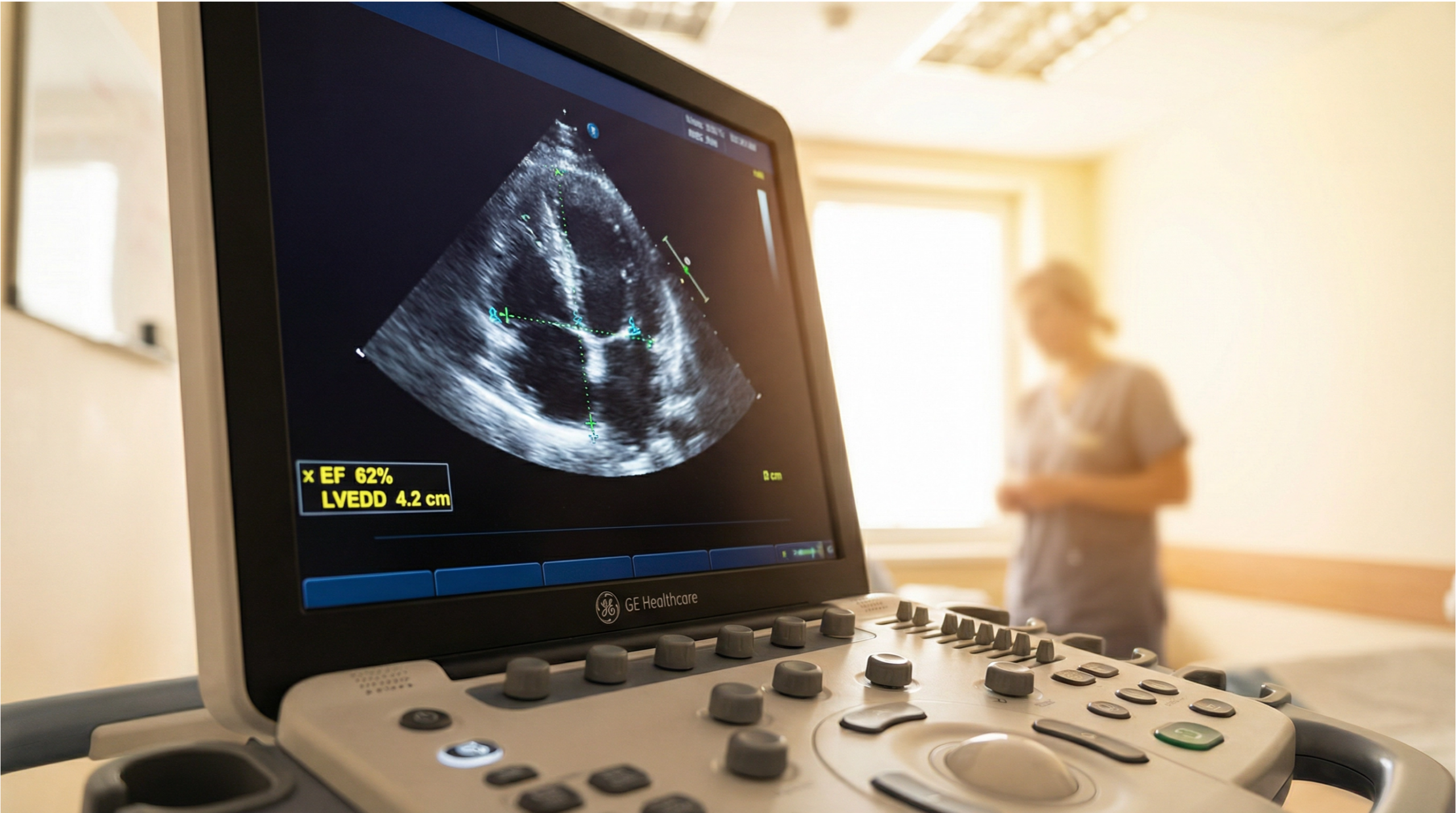
A standard echocardiogram (also called a transthoracic echo or TTE) is one of the safest heart tests available.
It uses ultrasound (sound waves), not radiation, and is generally well tolerated. This page explains what “safe” means in real life:
what you might feel, what the true risks are, which situations need a different pathway, and how to book with confidence.
If you have severe chest pain, collapse, severe breathlessness at rest, or stroke symptoms, seek urgent medical care first.
Private scanning is for clarity and next-step guidance — not emergency treatment.
Quick safety summary (plain English)
Most patients only need a standard transthoracic echo (TTE). It is non-invasive, uses ultrasound, and has a very strong safety profile.
The main “risks” are usually comfort-related, not medical.
What’s true for a standard TTE
Topic
What you should know
Radiation
None Ultrasound uses sound waves, not X-rays.
Pain
Usually no Most people feel gentle pressure; gel can feel cool.
Downtime
None You can usually drive and resume normal activities immediately after a standard TTE.
Medical risk
Very low For TTE, risks are minimal and uncommon.
When risk is higher
Other echo types Transoesophageal echo (TOE/TEE) and stress echo have different considerations.
If you want the full context (what echo checks, what it can’t show, and how it compares to ECG/CT), start here:
What is an ultrasound echo?
Safety is also about choosing the right pathway
The safest test is the test that answers your question without unnecessary risk.
If your symptoms are urgent, the safest pathway is urgent clinical assessment (not “waiting for a scan”).
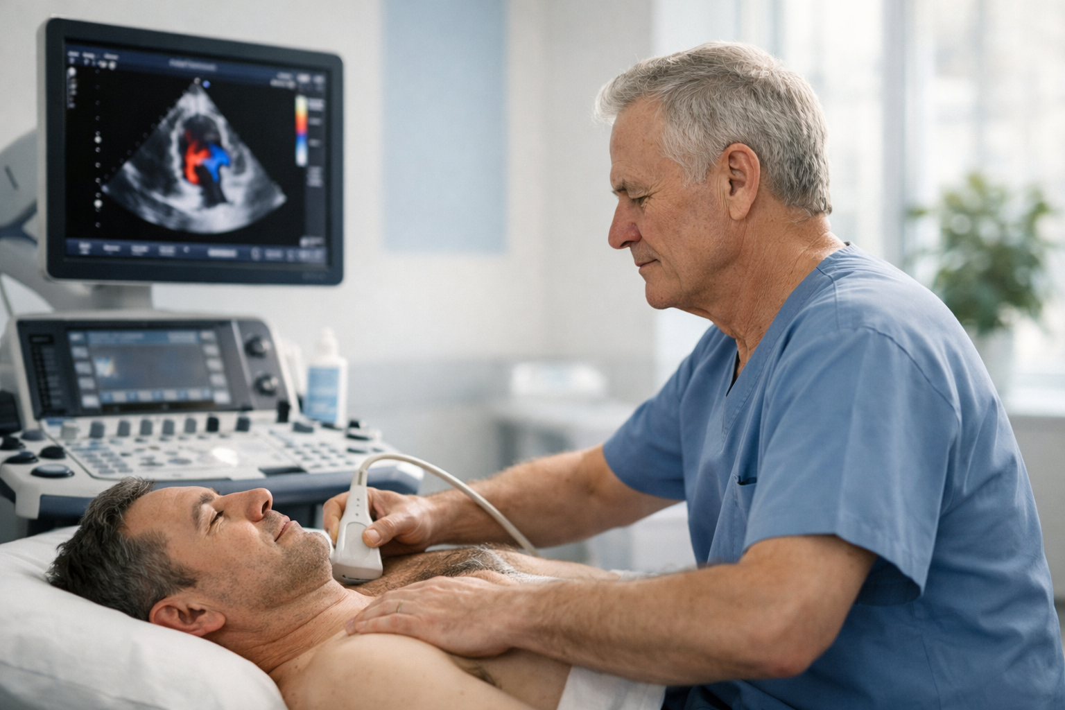
Most anxiety comes from uncertainty. Here’s what typically happens in a standard transthoracic echocardiogram.
Typical sensations
Cool gel on the chest at the start.
Gentle pressure as the probe is moved between rib spaces to obtain standard views.
You may be asked to change position or hold your breath briefly to improve image quality.
If your chest is tender
If you’ve had a recent injury, surgery, or you have pain in a specific spot, tell the clinician before the scan starts.
The exam can usually be adapted to keep things comfortable while still obtaining diagnostic views.
Real risks of a standard transthoracic echo (TTE)
For a standard TTE, serious complications are not expected. The realistic “risks” are typically minor and short-lived.
The bigger risk is booking the wrong test for your main symptom — which this cluster is designed to prevent.
What can happen (uncommon, usually mild)
Mild discomfort from probe pressure (especially if ribs/chest wall are sensitive).
Rare skin irritation from gel or wipes (tell us if you have known sensitivities).
Occasional lightheadedness from anxiety or breathing pattern changes (we pause, reset, and continue comfortably).
If you want the detailed “what it can and can’t tell you” boundary (accuracy limits, acoustic windows, and what echo doesn’t show),
use: Limitations of echocardiography.
No radiation doesn’t mean “no standards”
Ultrasound is used under established safety principles: scan for a justified reason, use appropriate settings, and keep exposure as low as reasonably achievable while obtaining diagnostic images.
This is one reason structured protocols and experienced operators matter.
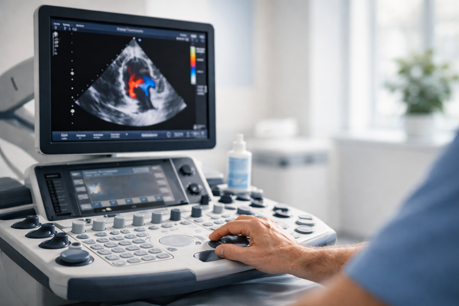
Other echo types (different comfort + risk profile)
Most people booking privately are having a standard transthoracic echo (TTE).
Less common echo types can be clinically appropriate in specific scenarios — but they have different preparation and risk considerations.
Echo type
Why it’s used
Comfort / risk notes
TTE (standard echo)
First-line assessment of valves, chambers, pumping function, pericardium and Doppler flow.
Very safe Non-invasive; usually no downtime.
TOE/TEE (transoesophageal echo)
Higher-detail views via an ultrasound probe in the oesophagus (often for specific structural questions).
Different pathway Can be uncomfortable; throat soreness is common afterwards; sedation may be used; rare throat injury is a recognised risk. (If your clinician requests this, preparation differs.)
Stress echo
Assesses heart function under stress (exercise or medication), in specific clinical pathways.
Clinician-led Risk relates to the stress component; suitability depends on history and supervision.
Practical point
If you were told you need TOE/TEE or stress echo specifically, tell us when booking so you’re directed to the right service and preparation steps.
Most private reassurance pathways begin with TTE.
Most people can have a standard TTE with no special precautions. These prompts simply help us keep the experience smooth and comfortable.
Tell us in advance if any of these apply
You have significant chest wall pain, a recent chest injury, or recent chest surgery.
You’ve had skin reactions to gels, adhesives, or wipes in the past (we can adapt products where possible).
You feel very anxious about the test (we can slow the process down and explain as we go).
You were specifically asked to have TOE/TEE or stress echo (different preparation and supervision).
Many people book because they want a focused, calm explanation. This is part of the Sonoworld approach: clarity you can act on.
How echo compares to other common heart tests (safety lens)
People often hear “ECG”, “echo”, “CT”, and assume they’re substitutes. They aren’t.
From a safety perspective, the right choice is about matching the test to the question — not doing more tests “just in case”.
Echo is a no-radiation test. CT uses X-rays (radiation) and may involve contrast dye in some pathways.
ECG has no radiation, but it measures electrical rhythm rather than structure.
Short, direct answers to the questions patients ask right before booking.
Is an echocardiogram safe?
A standard transthoracic echocardiogram (TTE) uses ultrasound (sound waves), not radiation, and is generally very safe and well tolerated.
The main issues are usually comfort-related rather than medical.
Does echo use radiation like CT?
No. Echo is ultrasound-based and does not use ionising radiation.
CT scans use X-rays (ionising radiation) and are used for different questions and pathways.
Can I drive home after a standard echo?
After a standard transthoracic echo (TTE), most people can return to normal activities immediately, including driving.
(If you were booked for a sedated procedure like TOE/TEE, driving restrictions can apply.)
Is it normal to feel pressure?
Yes. Gentle pressure is normal because the probe needs good contact between ribs to obtain clear images.
If anything feels uncomfortable, tell the clinician — the scan can usually be adjusted.
What are the risks of TOE/TEE compared to TTE?
TOE/TEE uses a probe in the oesophagus and can be uncomfortable; throat soreness afterwards is common, sedation may be used, and rare throat injury is a recognised risk.
This is a different pathway to standard TTE and is only used when clinically indicated.
What if I’m very anxious about the scan?
Tell us. Most anxiety improves when the process is explained step-by-step and you know what the test can (and can’t) show.
Many patients book privately specifically for calm clarity and structured reporting.