
- Home
- Women
- Men
- MSK
- Cardiovascular
- Screening
- About
- Book a Scan
- Blog
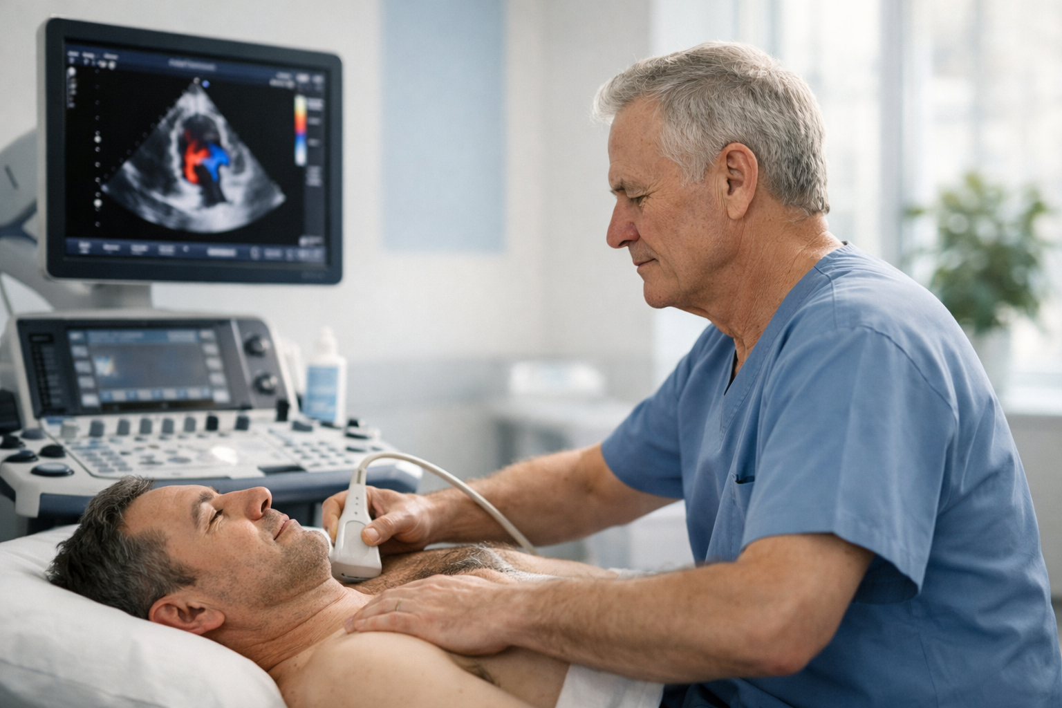
This is the most common confusion in cardiac testing: you have symptoms and you want the right test first. An ECG records the heart’s electrical activity (rhythm and conduction). An echo (echocardiogram) creates moving images of the heart’s structure and pumping function (valves, chambers, muscle, Doppler blood flow).
If you have severe chest pain, collapse, severe breathlessness at rest, or stroke symptoms, seek urgent medical care first. Private testing is best for clarity and next-step guidance — not emergency treatment.
You don’t need to memorise cardiology. Use this: ECG is the rhythm test. Echo is the structure/function test. Many real-world pathways use both — but usually in a deliberate order.
Palpitations, skipped beats, racing heart, episodic dizziness, fainting episodes, or “is my rhythm normal?” start with ECG (and often longer rhythm monitoring if episodes come and go).
Breathlessness on exertion, reduced exercise tolerance, a new murmur, valve concerns, or “is my heart pumping normally?” are echo questions.
Severe chest pain, collapse, severe breathlessness at rest, or stroke symptoms need urgent assessment (ECG + clinical pathway). Don’t use a private scan to replace emergency care.
This table is designed for patients: plain-English differences, plus what each test cannot do (so you don’t book the wrong thing).
| Feature | ECG (Electrocardiogram) | Echo (Echocardiogram / Heart ultrasound) |
|---|---|---|
| What it measures | Electrical activity: rhythm, heart rate, conduction pathways. | Moving images: chambers, valves, heart muscle function; Doppler blood flow. |
| Best for | Arrhythmia clues, conduction issues, acute changes (clinical context matters). | Murmur/valve assessment, pumping function, chamber size, pericardial fluid. |
| What it can miss | Intermittent rhythm issues if they don’t occur during the recording (may need monitoring). | Electrical rhythm diagnosis (echo is not rhythm capture) and direct coronary artery imaging. |
| How it feels | Electrodes on chest/limbs; you lie still; recording takes seconds. | Gel + probe on chest; gentle pressure; typical scan ~30–45 minutes. |
| Radiation | No | No |
| Typical next step | If symptoms persist: repeat ECG, Holter/event monitor, blood tests, clinical review. | If findings explain symptoms: management plan / GP / cardiology. If not: consider ECG/rhythm pathway or other imaging. |
ECG is the fastest way to answer “is my heart rhythm normal right now?” It’s often the first step in urgent pathways (especially chest pain), and a common first step for palpitations.
A normal ECG can still be “true” — it just means the rhythm was normal at that moment. If symptoms are episodic, clinicians often use longer monitoring (Holter/event monitor) to catch the episode. Echo may be added to check whether there’s a structural reason the rhythm is happening.

Echo answers: “How is my heart built, and how is it working?” It’s the main imaging test for murmurs/valves and a key test for breathlessness where a cardiac contribution is being considered.
ECG and echo are complementary: one is electrical, one is mechanical/structural. Here are the scenarios where clinicians commonly combine them.
ECG (or monitoring) answers “what rhythm is it?” Echo answers “is there an underlying structural contributor (valves, cardiomyopathy pattern, function)?”
Echo grades valve disease. ECG provides rhythm baseline and can show strain patterns that support the clinical picture.
Echo trends valve severity and function. ECG/monitoring checks rhythm evolution where clinically relevant.
In acute chest pain or collapse pathways, ECG is immediate. Echo may follow in hospital assessment depending on findings and clinical need.
Start with your “job to be done”: rhythm question → ECG. Structure/function question → echo. If you’re unsure, use the symptom guide and then choose the test that answers your main concern.
Symptoms that require an echoSonoworld provides private echocardiography (echo). ECG is commonly arranged via GP, cardiology, urgent care, or monitoring services depending on your symptom urgency and pattern.
Clinic: 29 Weymouth Street, Marylebone, London W1G 7DB • Call 020 3633 4902.

Read the “what is an ultrasound echo?” guide for the full map (echo vs ECG vs CT vs MRI), then decide based on the question you want answered.
What is an ultrasound echo? Limitations of echocardiographyThe questions people ask right before booking.
Clinic address: 29 Weymouth Street, Marylebone, London W1G 7DB.


