Written by Daniela Stan, Consultant Sonographer & Clinical Director, Sonoworld London.
Medically reviewed by Daniela Stan, Consultant Sonographer —
An armpit lump is most often a swollen lymph node reacting to a minor infection or a recent vaccination. Lipomas (soft fatty lumps) and cysts are the next most common causes. Most settle on their own within a few weeks. An ultrasound scan can tell you which one you have, usually in a single appointment.
In our Marylebone clinic, the great majority of armpit lumps we scan turn out to be exactly that: a benign, reactive lymph node. But a small number look different on the screen, and knowing what separates the two is the whole point of the scan.
When an armpit lump needs urgent attention
Most armpit lumps are not an emergency. A few are. These two lists are different, and mixing them up sends people to the wrong place.
Seek same-day medical review or go to A&E
These point to a spreading infection, which needs treating quickly. Call 999 or go to A&E if you have:
Redness or heat spreading outwards from the lump across the skin
Fever, shivering, or shaking chills
A painful swelling that is getting bigger over hours rather than days
Feeling very unwell, confused, or faint alongside the lump
See your GP promptly — within days, not weeks
These do not need an emergency department, but they do need a doctor soon, because they may lead to an urgent referral rather than a scan alone:
A lump that is hard, fixed in place, or does not move when you press it
A lump that keeps growing over weeks
Unexplained weight loss, drenching night sweats, or persistent tiredness alongside the lump
Dimpling, puckering, or an orange-peel texture in the skin of the armpit or nearby breast
A new change in the breast on the same side, or new nipple discharge
Any armpit lump you cannot explain that has not settled within three to four weeks
NICE guideline NG12 advises GPs to consider an urgent suspected-cancer referral — an appointment within two weeks — for an unexplained armpit lump in anyone aged 30 or over. Unexplained swollen glands alongside fever, night sweats, weight loss, or itching may prompt an urgent referral to investigate lymphoma. This page is general information and cannot replace an individual assessment by a clinician who can examine you.
What causes a lump in the armpit?
The armpit (the axilla) contains lymph nodes, fatty tissue, sweat glands, hair follicles, and in some people a small amount of breast tissue. A lump can come from any of them.
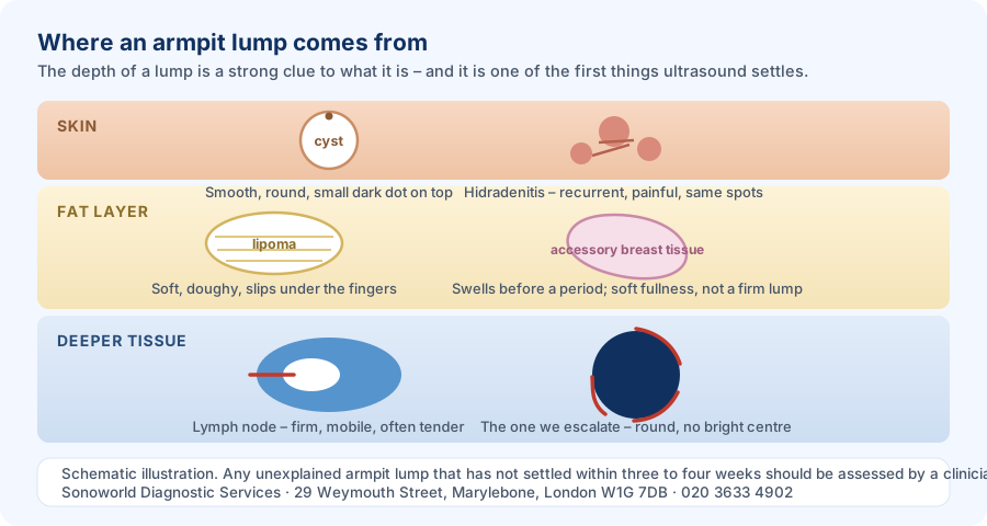
How deep a lump sits is one of the strongest clues to what it is, and it is one of the first things an ultrasound scan settles.
Reactive lymph nodes
This is the most common cause by a distance. Lymph nodes are small filters that swell when your immune system is working. A cut on the hand, a chest infection, a skin infection, a dental problem, or a recent jab in the arm can all make the nodes on that side swell. They are usually tender, move freely under the skin, and shrink back over two to four weeks.
Lipoma
A lipoma is a harmless lump of fat that sits just under the skin. It feels soft and doughy, slips away from your fingers when you press it, and is almost always painless. Lipomas grow very slowly, often over years, and usually need no treatment at all.
Epidermoid cyst (often called a sebaceous cyst)
A blocked hair follicle or skin gland can form a small sac filled with keratin, the same protein your skin and hair are made of. It feels smooth and round, sits within the skin rather than deep to it, and sometimes has a tiny dark dot on the surface. If it becomes infected it turns red, sore, and may discharge.
Hidradenitis suppurativa
This is a long-term skin condition affecting areas with sweat glands, including the armpits and groin. It causes painful lumps that come back in the same places, sometimes bursting and leaving scars or tunnels under the skin. It is commonly mistaken for repeated boils. It is treatable, and getting the right label on it early makes a real difference.
Accessory breast tissue
Some people are born with a small amount of extra breast tissue in the armpit. It often goes unnoticed until it responds to hormones — swelling and aching before a period, or during pregnancy and breastfeeding. It is entirely benign, and it is one of the causes most often missed, because it does not behave like a lump so much as a soft fullness that comes and goes.
Boils and abscesses
Shaving, waxing, and antiperspirants can irritate hair follicles, and an infected follicle can build into a boil or an abscess. These are hot, red, and genuinely painful. An abscess usually needs draining rather than antibiotics alone.
Less common causes
Occasionally an armpit lump is a swollen node containing cancer cells — from a breast cancer, a melanoma, or a lymphoma. This is far less common than the causes above, but it is the reason no unexplained lump should simply be ignored.
How to tell them apart
Feel is a useful clue, but it is only a clue. The table below shows the typical patterns. Plenty of lumps do not read the textbook, which is why imaging exists.
Reactive lymph node
Lipoma
Cyst
How it feels
Firm and rubbery, like a small bean
Soft and doughy
Smooth and round, sometimes squashy
Does it move?
Yes, freely
Yes — slips away under the fingers
Yes, but moves with the skin
Is it sore?
Often tender
Usually painless
Painless unless infected
How fast does it grow?
Appears over days, settles over weeks
Very slowly, over years
Slowly; can flare up suddenly
How deep is it?
Deeper, under the fat
Just under the skin
In the skin itself
Skin over it
Normal
Normal
May have a small central dot
What an ultrasound scan actually shows
Ultrasound is the right first test for a lump you can feel in the armpit. It uses sound waves, involves no radiation, needs no preparation, and shows soft tissue in real time. Crucially, it does something your fingers cannot: it looks inside the lump.
A normal lymph node
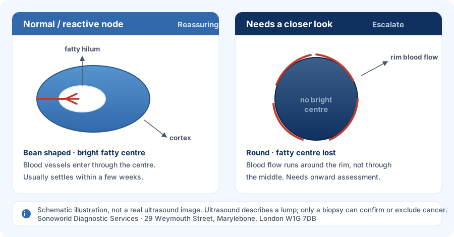
A healthy node has a recognisable shape on the screen. It is oval, like a kidney bean. It has a thin dark rim (the cortex) around a bright centre called the fatty hilum. Blood vessels enter neatly through that centre. A node that looks like this, in someone who has had a recent infection or vaccination, is reassuring.
A node that needs a closer look
Three changes matter. The node loses its bright fatty centre. It becomes round instead of bean-shaped. And blood flow appears around the rim of the node rather than entering through the middle. Any of these shifts the picture, and together they are the reason a scan gets escalated the same day.
The three changes our sonographers look for. Schematic illustration, not a real ultrasound image.
Lipomas and cysts on the screen
A lipoma appears as a well-defined, layered mass sitting in the fat, running parallel to the skin, with no fluid inside. A cyst appears as a dark, fluid-filled sac with a bright area behind it. These two look very different from each other and from a lymph node, which is why a scan can usually settle the question in one visit.
What ultrasound cannot do
Ultrasound describes a lump extremely well, but it cannot confirm or exclude cancer on its own. Only a tissue sample — a biopsy — can do that. What the scan does is decide, quickly and accurately, whether a biopsy is needed and how urgently. If your scan raises that question, you need onward specialist assessment, not reassurance from a website.
From our practice
When our sonographers scan an armpit lump at Weymouth Street, one thing decides what happens next. We are looking at whether the lymph node has kept its normal fatty hilum, whether it is still bean-shaped rather than round, and where the blood flow runs. The vast majority of the time the answer is straightforward — a benign, reactive node responding to a minor infection or a recent vaccination, and the patient leaves reassured the same morning.
When we do see those features change, we do not wait. We draft an urgent report immediately so the patient can be fast-tracked to their GP or directly to a specialist breast clinic for a biopsy. We chose that threshold deliberately: the loss of the fatty hilum, a round shape, and abnormal blood flow at the rim are the findings that change what a patient needs next, and a scan that spots them is only useful if the report moves as fast as the finding does.
That is a description of how we assess and escalate, not advice about your own lump. If you have found one, it needs looking at by a clinician who can examine you.
Armpit lumps after a vaccination
A swollen, tender node in the armpit on the same side as a recent jab is common and expected. Your immune system is doing precisely what the vaccine asked it to. It usually settles within a few weeks without any treatment.
Two practical points. If you are due a breast screening mammogram, mention a recent vaccination when you book, because the swelling can show up on the images. And if a node swollen after a vaccination is still there six weeks later, have it reassessed rather than assuming the jab is still the explanation.
NHS or private: which route is right for you?
NHS route
Sonoworld
How you get seen
GP appointment first, then referral
Book directly, no GP referral needed
Typical wait for a routine scan
Several weeks after referral
Same-day and next-day appointments
If red-flag features are present
Two-week-wait cancer pathway
Urgent report drafted for immediate onward referral
Results
Report to your GP, then a follow-up appointment
Instant verbal results; written report within 24 hours
Cost
Free
£235, all-inclusive
To be plain about it: if your GP has already put you on a two-week-wait pathway, stay on it. That route gives you the scan, the biopsy, and the specialist team together. Private imaging is most useful when you want an answer quickly, when a lump has been dismissed but has not gone away, or when the wait itself is the thing you cannot live with.
What happens at your appointment
Preparation: none. Eat, drink, and take your usual medication as normal.
What to wear: a top that is easy to remove or lift, so the armpit is accessible. A gown is provided.
What to bring: any previous scan reports, and a note of when you first noticed the lump and whether it has changed.
The scan: you lie down with your arm raised behind your head. Warm gel is applied and a small probe is moved over the area. It takes around 15 to 20 minutes and does not hurt.
Afterwards: your sonographer talks you through what they have seen before you leave. Your written report follows within 24 hours and can go directly to your GP or consultant.
You can book a lumps and bumps ultrasound scan for any lump you can feel. If the lump sits close to the breast, or you have noticed a breast change on the same side, a breast ultrasound may be the better test — call us and we will point you to the right one.
Frequently asked questions
Is an armpit lump usually cancer?
No. The great majority of armpit lumps are benign. Swollen lymph nodes responding to an infection or a recent vaccination are by far the most common cause, followed by lipomas and cysts. Cancer is an uncommon explanation. That said, no unexplained lump that persists beyond three to four weeks should be left unchecked, because the uncommon causes are the ones where early assessment matters most.
How long should I wait before getting an armpit lump checked?
If the lump appeared alongside an obvious infection or a recent jab and is settling, two to three weeks of watching is reasonable. If it has not gone within three to four weeks, is growing, is hard or fixed, or comes with weight loss or night sweats, get it assessed now rather than waiting. Anything with spreading redness or fever needs same-day medical review.
Can an ultrasound tell the difference between a lymph node, a cyst and a lipoma?
Yes, in most cases. These three have distinctly different appearances on ultrasound: a node has a bean shape and a bright fatty centre, a cyst is a dark fluid-filled sac, and a lipoma is a layered mass sitting within the fat. A scan can usually characterise the lump in a single appointment and say whether anything further is needed.
Can ultrasound rule out cancer completely?
No. Ultrasound is excellent at describing a lump and at identifying features that need further investigation, but only a biopsy can confirm or exclude cancer. The value of the scan is that it decides quickly and reliably whether a biopsy is needed, and how urgently. If a scan raises that question, you will need specialist assessment to answer it.
Why is my armpit lump painful?
Pain usually points towards inflammation or infection rather than anything sinister. Reactive lymph nodes are often tender. Infected cysts, boils, and abscesses can be very painful, with red, hot skin over them. A hard, painless, fixed lump is in some ways more concerning than a sore one, which is why pain alone is a poor guide to how worried to be.
Do I need a GP referral to get an armpit lump scanned at Sonoworld?
No. You can book a lumps and bumps ultrasound scan directly with us. If you already have a referral letter, bring it, because the clinical detail helps your sonographer focus the scan. If your GP has placed you on an urgent two-week-wait pathway, stay on that pathway as well as, not instead of, a private scan.
What does it mean if a lymph node has lost its fatty hilum?
The fatty hilum is the bright centre of a normal lymph node, where blood vessels enter. When it disappears, the internal architecture of the node has been replaced by something else — which can be infection, inflammation, or abnormal cells. On its own it is not a diagnosis. It is a finding that means the node should be assessed further, usually with a biopsy.
How much does an armpit lump ultrasound cost?
A lumps and bumps ultrasound scan at Sonoworld is £235. That price is all-inclusive: the scan itself, the consultant sonographer's time, instant verbal results at the end of the appointment, and a written report within 24 hours that can be sent to your GP or consultant. There are no separate reporting or booking fees.
Sonoworld is registered with the Care Quality Commission (CQC). Our sonographers are members of BMUS. All scans are performed at our Marylebone clinic: 29 Weymouth Street, London W1G 7DB.
Book Your Lumps and Bumps Ultrasound Scan at Sonoworld
Same-day and next-day appointments available at our Marylebone clinic. Instant verbal results. Written report within 24 hours. No GP referral required.