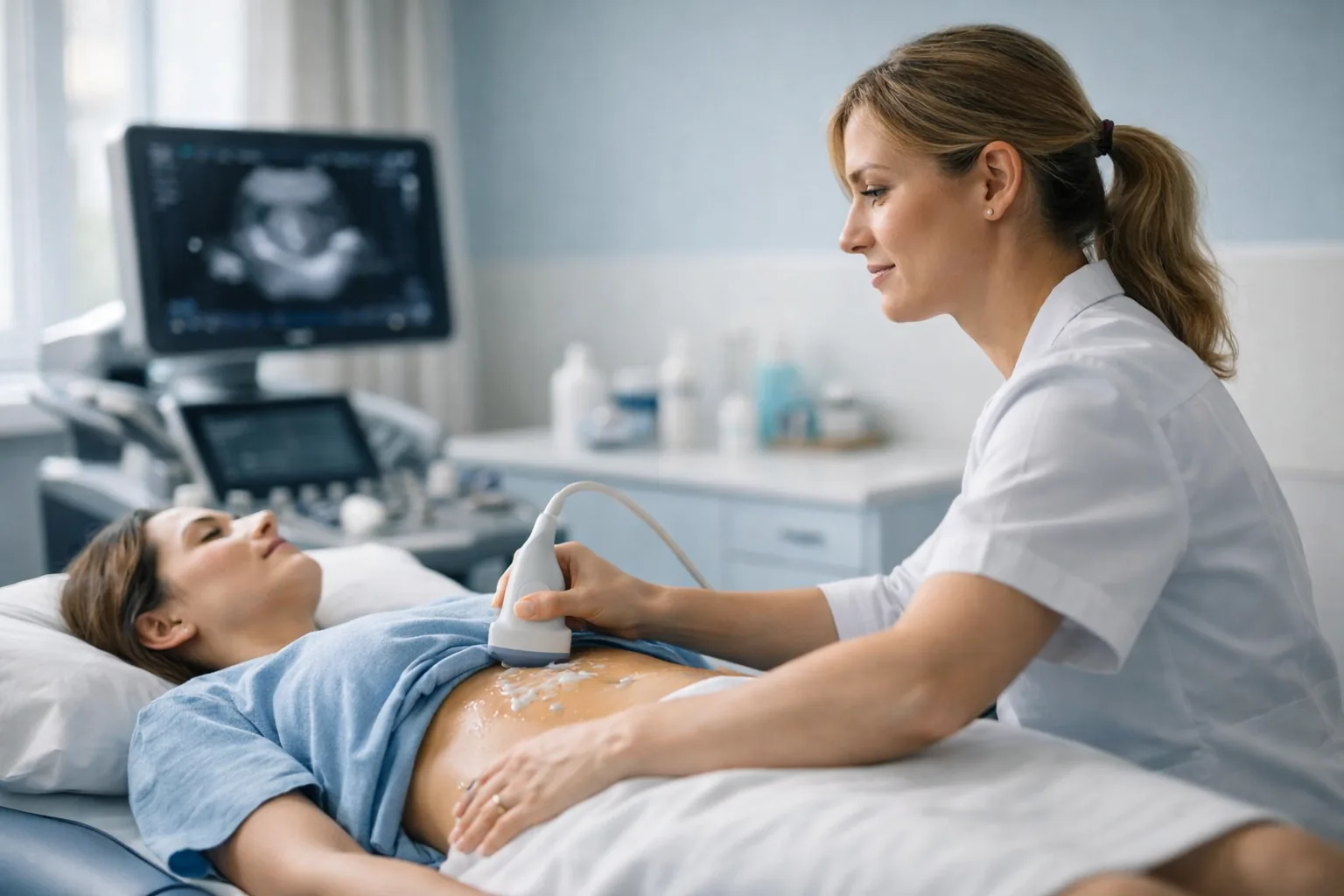
A pelvic ultrasound focused on uterine fibroids: how many there are, how big, exactly where they sit in the uterine wall, and whether they distort the cavity. Each fibroid is measured in three planes and classified using the FIGO system that gynaecologists use to plan treatment. Female consultant ultrasound practitioner, CQC-registered Marylebone clinic, same-day appointments, no GP referral required.
CQC Registered Good No GP Referral Needed FIGO Fibroid Mapping Marylebone, London
Booked as a pelvic ultrasound appointment. Instant verbal results. Written report within 24 hours.
£235All-inclusive price
20–30mScan duration
24hWritten report
CQCRated Good
About This Scan
Fibroid Mapping, Not Just Fibroid Detection
Fibroids (myomas) are benign growths of the muscle of the uterus and are very common: around one in three women develops them at some point. Many cause no trouble at all. Others cause heavy or prolonged periods, pelvic pressure, a need to pass urine often, back pain, or difficulty conceiving. Ultrasound is the first-line test recommended by NICE when fibroids are suspected or when heavy periods need investigating.
What makes a fibroid scan useful is the detail. Knowing that fibroids are present is rarely enough; your gynaecologist needs to know where each one sits. A fibroid that bulges into the cavity (submucosal, FIGO types 0 to 2) is the kind most likely to cause heavy bleeding and affect fertility, and may be removable through the cervix. A fibroid in the wall (intramural, types 3 to 5) or on the outer surface (subserosal, types 6 and 7) behaves differently and is treated differently. Your report maps every fibroid by size, position and FIGO type.
Ultrasound is accurate for most fibroids. When the uterus is very large or there are many fibroids, MRI may be recommended for mapping before surgery or embolisation, and your report will say so.
Why patients choose Sonoworld
CQC-registered clinic — inspected and rated Good.
Female consultant ultrasound practitioner with a gynaecological imaging focus.
Every fibroid measured in three planes and FIGO-classified.
Instant verbal results and a written report within 24 hours.
No GP referral required — self-refer online or by phone.
Transparent all-inclusive pricing at £235.
Who It Is For
When to Book a Fibroid Scan
Book this scan if you have symptoms that fibroids commonly cause, if fibroids have already been found and need measuring again, or if you are planning treatment or fertility care and need an accurate map.
Common reasons to book
Heavy periods: soaking through protection, passing clots, or bleeding for more than seven days.
Pelvic pressure, fullness or a firm swelling low in the abdomen.
Needing to pass urine frequently, or difficulty emptying the bladder.
Lower back pain or pain radiating to the legs without another cause.
Iron-deficiency anaemia attributed to heavy periods.
Difficulty conceiving or recurrent miscarriage.
Known fibroids that need monitoring for growth, or mapping before myomectomy, embolisation or hysterectomy.
What We Assess
What We Check During Your Scan
The whole pelvis is examined, with the fibroid map as the centrepiece of the report.
Fibroid Map
Number, size and FIGO position
Each fibroid is measured in three planes and classified as submucosal, intramural, subserosal or pedunculated. The dominant fibroid and the overall size of the uterus are recorded so that growth can be tracked on later scans.
Uterine Cavity
Cavity distortion, polyps and the endometrium
Whether any fibroid pushes into the cavity is the finding that matters most for bleeding and fertility. Endometrial thickness is measured and polyps, which cause similar bleeding, are looked for.
Ovaries
Ovarian and adnexal assessment
The ovaries are checked for cysts and other findings, and a fibroid on a stalk is distinguished from an ovarian mass using its blood supply and its connection to the uterus.
Blood Flow
Colour Doppler and degeneration
Colour Doppler shows the blood supply to each fibroid. Changes such as degeneration, calcification or cystic change are described, which helps explain pain and guides treatment choices.
Preparation
How to Prepare for Your Scan
Drink about a litre of water an hour before your appointment and do not empty your bladder: the transabdominal part of the scan needs a full bladder to see the whole uterus, which matters when fibroids are large. You will empty it before the transvaginal part. Continue any medication, including hormonal treatment, as normal.
On the day of your scan
Arrive 5 minutes before your appointment time.
Bring previous scan reports or gynaecology letters so fibroids can be compared over time.
Wear loose clothing that gives easy access to the lower abdomen.
No fasting is needed.
The transvaginal part is explained first and performed only with your consent.
You may bring a companion or chaperone.
The Process
What Happens at Your Fibroid Scan
1Clinical History
Your practitioner asks about your bleeding pattern, pain, pressure symptoms, previous scans and any planned treatment or fertility care.
2Transabdominal Scan
With a full bladder, the size and outline of the uterus and the overall distribution of fibroids are assessed from the outside.
3Transvaginal Scan
With your consent, the internal probe gives the detail needed to find small submucosal fibroids and to judge their relationship to the cavity.
4Fibroid Mapping
Each fibroid is measured, located and FIGO-classified; colour Doppler records its blood supply. The endometrium and ovaries are assessed in the same examination.
5Results and Written Report
Findings are explained straight away. A written report with measurements, FIGO types and images follows within 24 hours, ready for your GP, gynaecologist or fertility clinic.
Pricing
Scan Pricing
£235All-inclusive — no hidden fees
Consultant ultrasound practitioner examination
Transabdominal and transvaginal assessment with full fibroid mapping
Immediate verbal results
Written report with measurements and images within 24 hours
Report shared with your GP or specialist on request
The scan is booked as a pelvic ultrasound appointment; tell us that fibroids are the reason for the scan. Compare all women’s health examinations on the Women’s Health Scans Hub.
FAQs
Frequently Asked Questions
Can ultrasound detect all fibroids?
It detects the great majority, including small submucosal fibroids when the transvaginal probe is used. When the uterus is very large or there are many fibroids, some may be hidden behind others, and MRI is then the better mapping test before surgery. Your report will say if that applies to you.
What is the FIGO classification and why does it matter?
It is the international system that describes where a fibroid sits, from type 0 (entirely inside the cavity) to type 7 (on a stalk outside the uterus). The position predicts symptoms, fertility impact and which treatments are possible, which is why your gynaecologist will ask for it.
Do fibroids affect fertility?
Most do not. Fibroids that distort the cavity are the ones linked to difficulty conceiving and miscarriage, and removing them can help. The scan tells you which type you have.
How often should fibroids be monitored?
That depends on symptoms and size. Many gynaecologists repeat the scan after six to twelve months if a fibroid is large or symptoms are changing, and the measurements in your report make a direct comparison possible.
Is the transvaginal part necessary?
It is recommended because it finds small submucosal fibroids and shows the cavity clearly. It is explained first and performed only with your consent; a transabdominal-only scan can be done, with its limits noted in the report.
Do I need a GP referral?
No. You can self-refer and book online or by phone. The report can be shared with your GP or specialist afterwards.