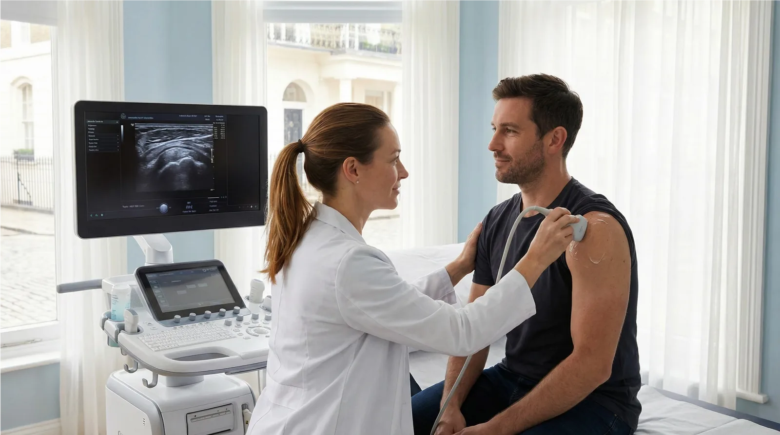
High-resolution, dynamic ultrasound of a torn or strained muscle: hamstring, calf, quadriceps, groin, biceps or any other muscle you can point to. The scan grades the tear, measures the gap and any bleeding, checks the tendon attachment and compares the injured side with the healthy one. Consultant sonographer, CQC-registered Marylebone clinic, same-day appointments, no GP referral required.
CQC Registered Good No GP Referral Needed Same-Day Results Marylebone, London
Book the MSK appointment for the area that is injured. Instant verbal results. Written report within 24 hours.
£235All-inclusive price
30mScan duration
24hWritten report
CQCRated Good
About This Scan
Grade the Injury Before You Plan the Recovery
A pulled hamstring, a calf that went during a sprint, a quadriceps or groin strain: muscle injuries are the most common injuries in sport, and the question that matters is always the same. How bad is it, and how long before I can train again? Ultrasound is the quickest way to answer the first part, and the answer shapes the second.
Muscle is an ideal tissue for ultrasound. The probe shows the fibres themselves, so the sonographer can see whether they are intact, partially torn or completely ruptured, measure the length of the tear and the size of any collection of blood, and locate the injury precisely: in the muscle belly, at the junction with the tendon, or at the tendon’s attachment to bone. Because the scan is dynamic, you can gently tense the muscle while it is imaged, which shows a gap that a still image would hide, and the other side is scanned for comparison.
Timing matters. The clearest pictures are obtained between about two days and a week after the injury, once bleeding has organised. Scanning within hours can underestimate the damage, so if you are in the first day or two we will advise on the best time to come. Ultrasound has limits too: deep muscles of the hip and pelvis, stress injuries of bone and complete tendon ruptures that need surgical planning are better assessed with MRI, and the report will say so when that applies.
Why patients choose Sonoworld
CQC-registered clinic — inspected and rated Good.
Consultant sonographers with specialist MSK expertise.
Dynamic imaging with side-to-side comparison, not a single still picture.
Instant verbal results and a written report within 24 hours.
No GP referral required — self-refer online or by phone.
Transparent all-inclusive pricing at £235.
Who It Is For
When to Book a Muscle Injury Scan
Book this scan after a sudden muscle injury during sport or exercise, for a muscle that keeps re-tearing, or for a lump or swelling in a muscle that has not settled.
Common reasons to book
A sudden pull, pop or tearing feeling in the hamstring, calf, thigh, groin or arm.
Bruising, swelling or a palpable gap or lump in a muscle.
Calf pain and swelling, where a tear needs to be told apart from a blood clot.
A strain that has not improved with rest and physiotherapy as expected.
Recurrent injury to the same muscle.
Follow-up of a known tear to check healing before returning to sport.
A firm lump within a muscle weeks after an injury.
What We Assess
What We Check During Your Scan
The injured muscle is examined along its whole length, from origin to insertion, at rest and under gentle contraction.
Tear Grade
Strain, partial tear or complete rupture
Grade 1 injuries show swollen but intact fibres; grade 2 a partial tear with a measurable gap; grade 3 a complete rupture with the muscle ends pulled apart. The length and cross-sectional extent of the tear are recorded.
Location
Belly, musculotendinous junction or tendon
Where the tear sits predicts how it heals. Injuries at the junction with the tendon and avulsions where the tendon pulls off the bone (proximal hamstring, distal biceps, Achilles) are identified because they may need a specialist opinion.
Haematoma
Bleeding and fluid collections
A collection of blood within or between muscles is measured and described. Its size influences rehabilitation and whether drainage should be discussed.
Healing and Complications
Scar tissue, calcification and the veins
On follow-up scans, scar formation and healing are assessed. Calcification within a healing muscle (myositis ossificans) is identified, and for calf injuries the deep veins can be checked in the same visit when a clot is a concern.
Preparation
How to Prepare for Your Scan
No preparation is needed. Wear or bring shorts, or clothing that can be moved away from the injured area. If the injury happened in the last 48 hours, call us: we will book you for the time when the scan is most informative. Note the date and mechanism of the injury, as both help with interpretation.
On the day of your scan
Arrive 5 minutes before your appointment time.
Bring any previous scan reports, MRI results or physiotherapy notes.
Wear comfortable, loose clothing for the area being scanned.
No fasting or special preparation is required.
You may be asked to gently tense or move the muscle during the scan.
You may bring a companion to your appointment.
The Process
What Happens at Your Muscle Injury Scan
1Clinical History
Your sonographer asks how and when the injury happened, where it hurts and what you have been able to do since. You will be asked to point to the exact spot.
2Dynamic Ultrasound Scan
Gel is applied and the whole muscle is scanned from origin to insertion at rest and under gentle contraction, with the opposite side imaged for comparison.
3Measurement and Grading
The tear is graded and measured, any haematoma is sized, and the tendon attachments are checked for avulsion.
4Written Report Within 24 Hours
Findings are explained straight away, and a written report with images follows within 24 hours for your physiotherapist, GP or sports physician to plan rehabilitation and return to sport.
Pricing
Scan Pricing
£235All-inclusive — no hidden fees
Consultant sonographer examination
Dynamic real-time imaging with side-to-side comparison
Immediate verbal results
Comprehensive written report within 24 hours
Report shared with your physiotherapist, GP or specialist on request
Book the MSK appointment for the region of the injury (for example hip for a hamstring or groin strain, knee for the thigh, ankle for the calf and Achilles). View all MSK scan types on the MSK Ultrasound Scans Hub.
FAQs
Frequently Asked Questions
How soon after the injury should I have the scan?
Ideally between two days and a week after the injury. By then bleeding has organised and a tear and its size are at their clearest. A scan within the first day or two can underestimate the injury, so if you are very early we will suggest the best time.
Ultrasound or MRI for a muscle tear?
For most hamstring, calf, quadriceps and groin strains ultrasound answers the question quickly, dynamically and at lower cost. MRI is better for deep muscles, for bone and stress injuries, and for complete ruptures that may need surgery. If your scan suggests MRI would add something, the report will say so.
Can the scan tell me how long I will be out of sport?
It gives the facts the timeline is built on: the grade, the exact location and the size of the tear and any haematoma. Your physiotherapist or sports physician uses those to set the rehabilitation plan. Higher grades and injuries at the tendon junction generally take longer.
My calf is painful and swollen. Is it a tear or a clot?
Both can look and feel similar, and both can be assessed with ultrasound. Tell us when booking and the deep veins can be examined in the same visit. If you also have breathlessness or chest pain, seek urgent medical care first.
Which appointment do I book?
Book the MSK appointment for the region of the injury: hip for the hamstring or groin, knee for the thigh, ankle for the calf and Achilles, shoulder for the upper arm. If you are unsure, call 020 3633 4902 and we will book it for you.
Do I need a GP referral?
No. At Sonoworld you can self-refer directly without a GP referral. Simply book online or call our team.