Expert ankle ultrasound imaging for Achilles tendinopathy, ankle ligament sprains, peroneal tendon tears, and plantar fasciitis. CQC-registered Harley Street clinic, same-day appointments, no GP referral required.
Instant verbal results. Written report within 24 hours.

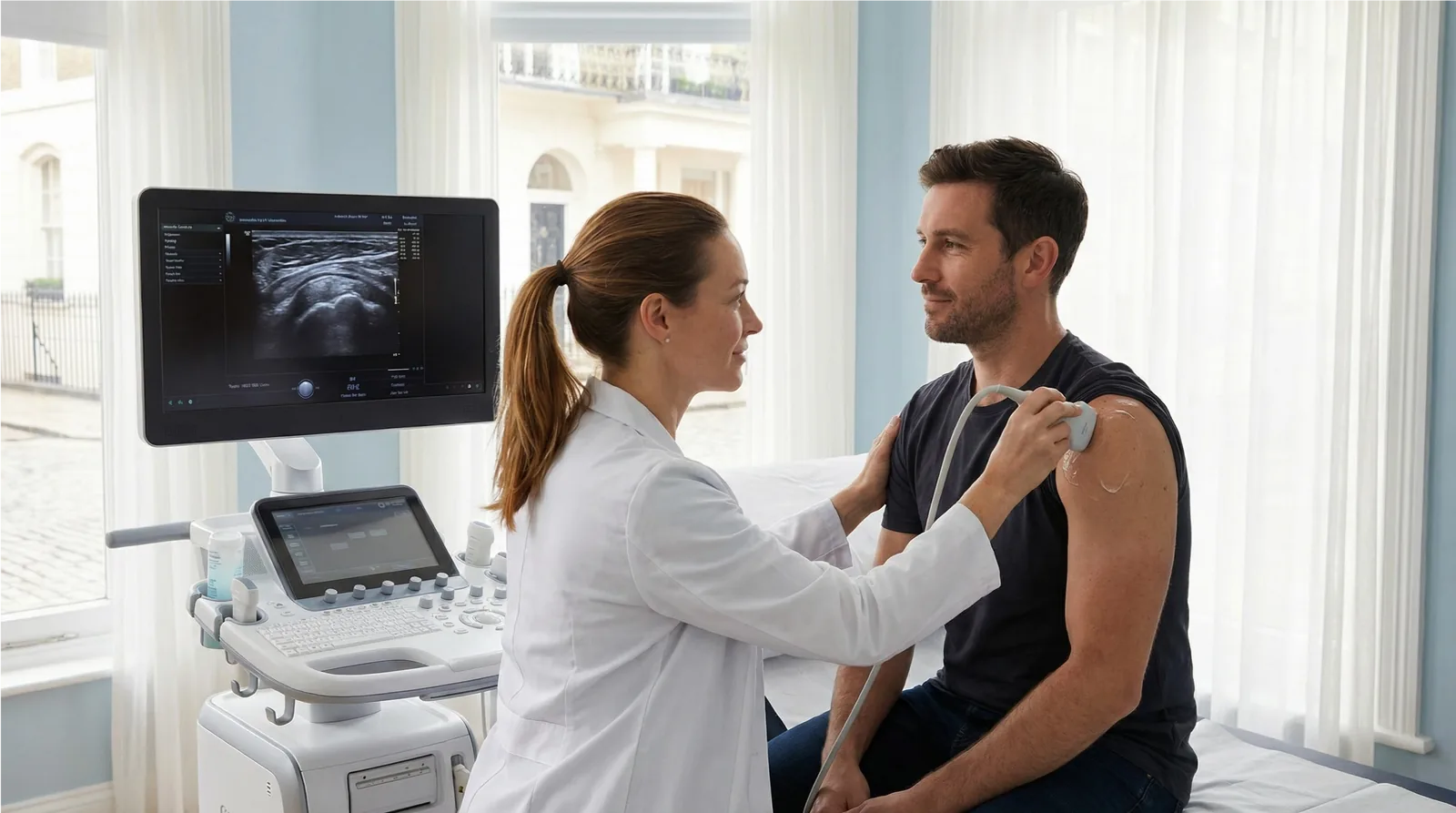
Musculoskeletal (MSK) ultrasound provides real-time, high-resolution imaging of joints, tendons, ligaments, and soft tissue. Unlike MRI, it allows dynamic assessment — structures can be examined in motion, both sides compared simultaneously, and the sonographer can provide immediate verbal feedback during the examination.
At Sonoworld, all MSK scans are performed by HCPC-registered consultant sonographers with specialist musculoskeletal expertise, using high-resolution GE ultrasound equipment. No GP referral is required, and a comprehensive written report is delivered within 24 hours.
Our consultant sonographers perform a systematic assessment of all relevant structures. Here is what is included in your examination.
Detailed assessment of the Achilles tendon throughout its length for tendinopathy, partial tears, and insertional enthesopathy. The retrocalcaneal bursa is also assessed. Achilles tendinopathy is one of the most common MSK conditions presenting to our clinic.
Evaluation of the anterior talofibular ligament (ATFL), calcaneofibular ligament (CFL), and medial ligament complex for sprains and tears. Dynamic stress testing can be performed during the scan to assess ligament laxity.
Assessment of the peroneal tendons for tears, tenosynovitis, and subluxation. The plantar fascia is assessed at its calcaneal origin for plantar fasciitis and partial tears.
No preparation is required for an ankle ultrasound scan. Wear or bring socks and shoes that can be easily removed. You may be asked to move your ankle during the scan for dynamic assessment of tendon subluxation or ligament laxity.
Arrive at our Harley Street clinic. Our team will take a brief clinical history, including your symptoms, duration, and any previous investigations or treatment.
Gel is applied to the skin and your consultant sonographer scans the affected area using a high-resolution probe. You may be asked to move the joint during the scan for dynamic assessment.
Your sonographer explains their findings immediately after the scan, answering any questions about what was seen and what it means for your management.
A comprehensive written report is emailed to you within 24 hours. You may share this directly with your GP, physiotherapist, or specialist.
View all MSK scan types on the MSK Ultrasound Scans Hub.
An ankle ultrasound scan provides detailed imaging of the Achilles tendon, ankle ligaments, peroneal tendons, plantar fascia, and ankle joint. It can identify Achilles tendinopathy, partial tendon tears, ankle ligament sprains, peroneal tendon tears, plantar fasciitis, and joint effusion.
Achilles tendinopathy is degeneration of the Achilles tendon, causing pain and stiffness at the back of the heel. Ultrasound can confirm the diagnosis, assess severity, and guide treatment decisions including shockwave therapy or PRP injection.
No. At Sonoworld, you can self-refer directly without a GP referral. Simply book online or call our team.
The scan typically takes 20–30 minutes. Your sonographer will provide immediate verbal feedback, and a written report will be emailed within 24 hours.
Yes. Ultrasound is the preferred investigation for plantar fasciitis. It can confirm the diagnosis by measuring plantar fascia thickness at the calcaneal origin (>4mm is considered abnormal) and guide injection if required.
If imaging confirms inflammation or soft-tissue injury, Sonoworld also offers ultrasound-guided cortisone injection for ankle pain — a precise, same-day treatment performed under real-time ultrasound guidance.
If your ankle scan reveals inflammation or a painful soft tissue condition, you may benefit from an ultrasound-guided cortisone injection for ankle pain at Sonoworld. Our precision-guided injections are performed under real-time ultrasound imaging for accurate placement and faster relief.