Aortic Stenosis: Causes, Diagnosis, and Treatment Options
Aortic stenosis is the most common valvular heart disease in the UK, affecting a significant proportion of the older population. It occurs when the heart's aortic valve narrows, restricting blood flow from the heart to the rest of the body. This comprehensive guide explains the causes, symptoms, and how an echocardiogram is used to accurately grade the severity of the condition.
CQC-registered clinic
Same-day appointments
No GP referral needed
Report within 24 hours
The aortic valve is the final gateway between the heart and the rest of the body. When the heart's left ventricle contracts, the aortic valve opens to allow oxygen-rich blood to flow into the aorta (the main artery). Aortic stenosis occurs when the leaflets of this valve become stiff, thick, or fused together, narrowing the opening.
This narrowing forces the heart muscle to work significantly harder to pump blood through the restricted valve. Over time, this increased workload causes the heart muscle to thicken (left ventricular hypertrophy) and eventually weaken, leading to heart failure if left untreated. A large-scale study of nearly 30,000 echocardiograms found that 7.2% of patients had some degree of aortic stenosis, with the prevalence rising sharply in those over 75 years of age [1].
Primary Causes
Age-related calcification: The most common cause. Over decades, calcium deposits build up on the valve leaflets, making them stiff. This degenerative process accounts for over 93% of cases in developed countries [1].
Bicuspid aortic valve: A congenital condition where the valve has only two leaflets instead of the normal three. This abnormal structure leads to earlier wear and tear, often causing stenosis in patients in their 40s or 50s.
Rheumatic fever: A complication of untreated strep throat that can cause scar tissue to form on the aortic valve. While now rare in the UK (accounting for only about 3% of cases), it remains a significant cause globally [1].
The Silent Progression
Aortic stenosis is often described as a "silent" disease because it progresses slowly over many years. The heart compensates for the narrowing valve by thickening its muscle walls. During this compensatory phase, patients may feel completely normal and remain asymptomatic.
However, once symptoms do appear, the condition has typically reached a severe stage, and the prognosis without intervention declines rapidly. This highlights the importance of early detection through an echocardiogram, especially if a doctor detects a heart murmur during a routine stethoscope examination.
Symptoms of aortic stenosis
The onset of symptoms in aortic stenosis is a critical clinical milestone. Research shows that the severity of presenting symptoms directly correlates with long-term survival outcomes, even after the valve is replaced [2].
When to seek emergency care
If you experience sudden, severe chest pain, fainting (syncope), or extreme difficulty breathing, call 999 immediately. These can be signs of critical aortic stenosis or a heart attack.
Common
Breathlessness (Dyspnoea)
Initially noticed only during physical exertion, such as climbing stairs. As the condition worsens, shortness of breath may occur at rest or when lying down. This is often a sign that fluid is backing up into the lungs (heart failure).
Warning Sign
Chest Pain (Angina)
A feeling of tightness, pressure, or squeezing in the chest, particularly during activity. This occurs because the thickened heart muscle requires more oxygen than the narrowed coronary arteries can supply.
Severe
Fainting (Syncope)
Feeling dizzy, lightheaded, or completely losing consciousness, especially during or immediately after physical exertion. This happens when the narrowed valve prevents the heart from pumping enough blood to the brain.
General
Fatigue and Weakness
A profound sense of tiredness and a noticeable decline in the ability to perform normal daily activities. Patients often subconsciously reduce their activity levels to avoid triggering other symptoms.
Diagnosis and grading
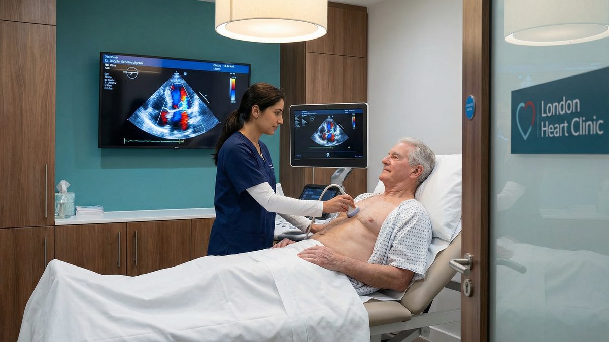
An echocardiogram is the primary and most important diagnostic tool for aortic stenosis. This specialised ultrasound-scan allows cardiologists to directly visualise the valve, measure the severity of the narrowing, and assess how well the heart muscle is coping with the increased workload.
Key Echocardiogram Measurements
During the scan, the sonographer will use Doppler ultrasound to measure three critical parameters that determine the severity of the stenosis:
Aortic Valve Area (AVA): The physical size of the valve opening. A normal valve area is 3.0 to 4.0 cm². Severe stenosis is defined as an area less than 1.0 cm² [3].
Peak Jet Velocity (Vmax): The maximum speed at which blood is forced through the narrowed valve. A velocity greater than 4.0 metres per second indicates severe stenosis.
Mean Pressure Gradient: The difference in pressure between the left ventricle and the aorta. A mean gradient greater than 40 mmHg is a marker of severe disease.
The Diagnostic Challenge: Low-Gradient AS
Grading aortic stenosis is not always straightforward. In approximately 25% of patients with severe stenosis, the pressure gradient appears deceptively low (under 40 mmHg) despite a critically narrowed valve area (under 1.0 cm²) [1].
This condition, known as Low-Flow, Low-Gradient (LFLG) severe aortic stenosis, occurs when the heart muscle has become too weak to generate a high pressure gradient. These patients represent a diagnostic challenge and often require advanced echocardiographic techniques or a cardiac CT scan to confirm the severity of the calcification and ensure they receive timely treatment [4].
Treatment options: TAVI vs SAVR
There are no medications that can reverse or slow the progression of aortic valve narrowing. Once severe aortic stenosis becomes symptomatic, the only effective treatment is to replace the diseased valve. Without valve replacement, the prognosis is poor, with a high mortality rate within two to three years of symptom onset.
Procedure
How it works
Recovery Time
Best suited for
TAVI (Transcatheter Aortic Valve Implantation)
A minimally invasive procedure. A new valve is delivered via a catheter, usually inserted through an artery in the groin, and expanded inside the old, diseased valve.
Typically 2 to 5 days in hospital. Faster return to normal activities.
Patients of all surgical risk levels. Recent landmark trials have proven TAVI is non-inferior to open surgery even in low-risk patients [5][6].
SAVR (Surgical Aortic Valve Replacement)
Traditional open-heart surgery. The chest is opened, the patient is placed on a bypass machine, the diseased valve is removed, and a new mechanical or tissue valve is sewn in.
Typically 7 to 10 days in hospital. Several weeks for full sternum healing.
Younger patients, those needing a mechanical valve, or patients requiring other simultaneous heart surgeries (e.g., coronary bypass).
Recent Clinical Evidence: The landmark DEDICATE-DZHK6 trial (published in the New England Journal of Medicine in 2024) compared TAVI and SAVR in 1,414 low-to-intermediate risk patients. The study found that TAVI was non-inferior to open surgery, with a lower incidence of death or stroke at 1 year (5.4% for TAVI vs 10.0% for SAVR) [5]. Additionally, 8-year follow-up data from the NOTION trial confirms that transcatheter valves demonstrate excellent long-term durability [7].
Frequently asked questions
Can aortic stenosis be cured with medication?
No. Aortic stenosis is a mechanical problem caused by physical narrowing and calcification of the valve. While medications can help manage symptoms or treat concurrent conditions like high blood pressure, they cannot reverse the stenosis. Valve replacement (TAVI or SAVR) is the only definitive treatment.
How often should I have an echocardiogram if I have mild aortic stenosis?
Clinical guidelines generally recommend an echocardiogram every 3 to 5 years for mild aortic stenosis, every 1 to 2 years for moderate stenosis, and every 6 to 12 months for severe asymptomatic stenosis. However, if you develop new symptoms at any time, you should arrange a scan immediately.
What is the difference between aortic stenosis and aortic sclerosis?
Aortic sclerosis is the early stage of the disease process, where the valve leaflets are thickened and calcified but the blood flow is not yet significantly restricted. Aortic stenosis occurs when the calcification progresses to the point where it obstructs the flow of blood out of the heart.
Do I need a GP referral to book an echocardiogram?
No. At Sonoworld, you can self-refer and book a private echocardiogram directly. This allows you to bypass waiting lists and get immediate clarity on your heart health. Read more about understanding your report.
References
Ramos J, et al. (2018). Large-scale assessment of aortic stenosis: facing the next cardiac epidemic? European Heart Journal - Cardiovascular Imaging.
Ben-Shoshan J, et al. (2019). Relation of Clinical Presentation of Aortic Stenosis and Survival Following Transcatheter Aortic Valve Implantation. The American Journal of Cardiology.
Minners J, et al. (2008). Inconsistencies of echocardiographic criteria for the grading of aortic valve stenosis. European Heart Journal.
Bismee N, et al. (2025). Discordant High-Gradient Aortic Stenosis: A Systematic Review. Journal of Cardiovascular Development and Disease.
Blankenberg S, et al. (2024). Transcatheter or Surgical Treatment of Aortic-Valve Stenosis. The New England Journal of Medicine.
Toff W, et al. (2022). Effect of Transcatheter Aortic Valve Implantation vs Surgical Aortic Valve Replacement on All-Cause Mortality in Patients With Aortic Stenosis: A Randomized Clinical Trial. JAMA.
Jørgensen T, et al. (2021). Eight-year outcomes for patients with aortic valve stenosis at low surgical risk randomized to transcatheter vs. surgical aortic valve replacement. European Heart Journal.