
Home › Patient Guides › Cardiovascular Health
Leg Swelling and Calf Pain: Could It Be a DVT?
Most swollen legs are not a blood clot. But swelling in one leg that comes on over hours or days — especially with calf pain, warmth or a change in skin colour — is the pattern that needs same-day medical assessment, not a scan booked for next week. This guide explains how to tell an urgent picture from a routine one, what a DVT scan actually looks for, and when a private scan is and is not the right route.
When leg symptoms need urgent care, not a booked scan
- Breathlessness, chest pain, coughing up blood, or feeling faint — call 999. These can indicate a pulmonary embolism, where a clot has travelled to the lungs. This is a medical emergency regardless of how the leg looks.
- Swelling in one leg with pain, warmth, or redness coming on over hours or days — contact your GP, NHS 111, or an urgent treatment centre for same-day assessment. Suspected DVT is assessed and treated on an urgent pathway, and that pathway starts with a clinical assessment, not an imaging booking.
- A leg that is cold, pale, numb, or that you cannot move — go to A&E. This is a different emergency from DVT and needs immediate assessment.
- Swelling with fever, spreading redness, or feeling generally unwell — seek same-day medical review; this may be a skin or soft-tissue infection.
Private imaging is for clarity and next-step guidance. It is not a substitute for urgent assessment, and it should never delay it.
On this page
One leg or two? The first question that matters
It sounds almost too simple, but whether one leg or both legs are affected changes the likely explanation more than any other single feature.
Both legs swollen, roughly equally — this pattern usually points away from a clot in a single vein. Swelling on both sides is more commonly related to prolonged standing or sitting, heat, pregnancy, certain medications, or conditions affecting the heart, kidneys, liver or thyroid. It still deserves a medical opinion if it is new, persistent or worsening, but it is less characteristic of a DVT.
One leg swollen, the other normal — this asymmetry is what raises the question of a deep vein thrombosis. A clot in a deep vein obstructs the return of blood from that limb, so the swelling is confined to the affected side. Add calf pain, warmth, or a change in colour, and the clinical suspicion rises further.
Asymmetry alone does not confirm a clot. A ruptured Baker's cyst, a calf muscle tear, cellulitis, or a long-standing venous problem can all swell one leg. That is precisely why suspected DVT is assessed clinically first: the examination, your risk factors and a blood test together determine whether imaging is needed and how urgently.
What a DVT typically feels like
Deep vein thrombosis has no single presentation, and a meaningful proportion of clots cause few symptoms at all. Where symptoms do occur, the commonly reported pattern is:
- Swelling of the calf, ankle or whole leg on one side
- An aching, cramping or tight pain, often in the calf, that is worse on standing or walking
- Warmth over the affected area
- Skin that looks red, darkened or discoloured compared with the other leg
- Veins near the surface that appear more prominent than usual
Two cautions matter here. First, the absence of these features does not exclude a clot. Second, their presence does not confirm one — several harmless conditions produce an almost identical picture. Neither this list nor any online checklist can replace clinical assessment, which is why the guidance above routes suspected DVT to same-day review rather than to a scan booking.
A note on calf pain without swelling. Isolated calf pain after unaccustomed exercise, with no swelling, no warmth and no colour change, is far more often muscular than vascular. It still warrants review if it persists, worsens, or appears without an obvious cause — particularly after recent surgery, immobility or long-distance travel.
Other common causes of a swollen leg
Understanding the alternatives helps explain why assessment comes before imaging.
- Chronic venous insufficiency. Valves in the leg veins stop closing properly, so blood pools. Swelling is typically worse by the end of the day and better after a night's rest, and is often accompanied by varicose veins or skin changes around the ankle.
- Baker's cyst. A fluid-filled swelling behind the knee. If it ruptures, fluid tracks into the calf and produces pain and swelling that can closely mimic a DVT.
- Muscle or tendon injury. A calf tear causes sudden pain, often with a memorable moment of onset, and bruising may appear later.
- Cellulitis. A bacterial skin infection producing redness, heat, tenderness and often fever. Needs antibiotics, not anticoagulation.
- Lymphoedema. Impaired lymphatic drainage, sometimes following surgery or radiotherapy, producing persistent swelling that is characteristically firmer.
- Medication effects. Some blood-pressure medicines, steroids and hormonal treatments cause fluid retention, usually affecting both legs.
What raises your risk
Risk factors matter because they change how seriously a given set of symptoms is taken. Recognised contributors include recent surgery — particularly hip, knee or abdominal — prolonged immobility including long-haul travel, pregnancy and the weeks following delivery, combined hormonal contraception and hormone replacement therapy, active cancer and some of its treatments, a previous DVT or pulmonary embolism, an inherited clotting tendency, obesity, smoking, and increasing age.
Having risk factors does not mean you have a clot, and having none does not mean you do not. They inform clinical judgement rather than replace it.
From our practice
Occasionally a scan booked for reassurance raises suspicion of an acute clot during the appointment itself. Our protocol is that the situation is explained to the patient there and then, rather than left to the written report.
What follows depends on severity. In most cases the patient is asked to attend their nearest emergency department, taking the report with them, so that treatment can be started without the findings having to be established a second time. Where the presentation warrants it, an ambulance is called from the clinic.
This is why we would rather you were assessed urgently elsewhere first if a clot is suspected: our part is the imaging, and the treatment decision belongs on an urgent pathway.
What a DVT ultrasound scan involves
A deep vein thrombosis scan is a duplex ultrasound: it combines standard greyscale imaging with Doppler, which shows blood flow direction and speed.
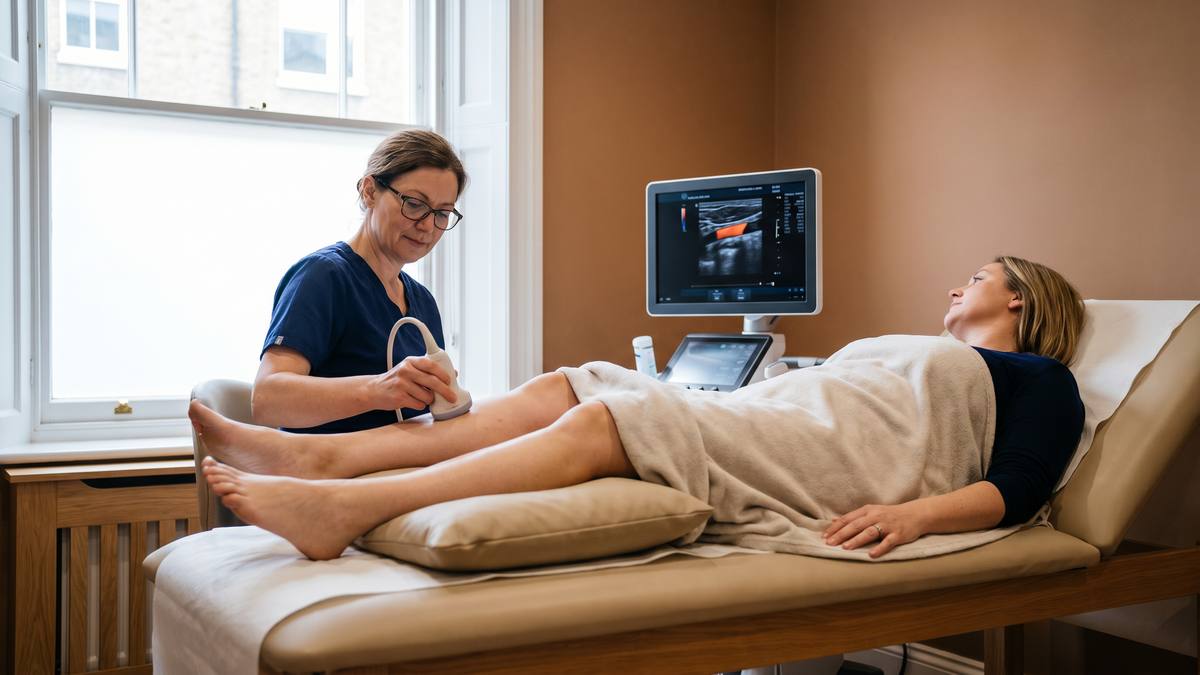
The examination is straightforward and non-invasive. You lie on the couch with the leg exposed and slightly rotated outwards. Warmed gel is applied and a probe is passed along the course of the deep veins, from the groin down through the thigh and into the calf. The sonographer applies gentle pressure at intervals — a healthy vein flattens completely under pressure, whereas a vein containing clot does not. That compression test is the core of the examination. Colour Doppler then demonstrates whether blood is flowing normally through the vessel, and spectral waveforms show how flow responds to breathing and to gentle squeezing of the calf.
The scan takes approximately 30 minutes. No preparation is needed: you can eat, drink and take your usual medication as normal, and no GP referral is required. You will be asked to undress from the waist down, and a chaperone is available on request.
At Sonoworld a DVT ultrasound scan costs £235. You receive instant verbal results at the appointment, with your written report following within 24 hours.
NHS and private routes compared
This is where the distinction genuinely matters, and where a private clinic ought to be candid.
If a DVT is suspected acutely, the NHS urgent pathway is the right route. Suspected DVT is assessed using a structured clinical scoring system alongside a D-dimer blood test, and where imaging is indicated it is arranged within a defined urgent timeframe. Crucially, that pathway also provides the thing imaging alone cannot: immediate access to anticoagulation if a clot is confirmed, and follow-up to establish why it happened. A private scan gives you a picture; it does not give you a treatment pathway.
Private imaging is better suited to different questions. It is a reasonable route when acute DVT has already been excluded or considered unlikely and you want to understand persistent swelling; when you are investigating chronic venous problems such as varicose veins or venous insufficiency; when you want surveillance after a previously treated clot; or when you have been advised imaging is appropriate but not urgent, and waiting is causing you difficulty.
If a scan performed here raises suspicion of an acute clot, we will tell you at the appointment and direct you to urgent care the same day. We would rather send you elsewhere quickly than hold on to a finding that needs treating within hours.
Frequently Asked Questions
Can I book a private scan instead of going to A&E?
Not if a DVT is suspected acutely. Suspected DVT needs same-day clinical assessment, because the decision that matters most is whether to start anticoagulation, and that decision follows clinical scoring and blood tests as well as imaging. Book a private scan when acute clot has been excluded or considered unlikely and the outstanding question is about chronic swelling, venous insufficiency, or surveillance after a previous clot.
How accurate is ultrasound for detecting a DVT?
Compression duplex ultrasound is the established first-line imaging test for DVT in the legs and performs well for clots above the knee. Clots confined to the smaller calf veins are more difficult to demonstrate reliably, which is one reason a repeat scan is sometimes recommended after an interval, and why imaging is interpreted alongside clinical assessment rather than in isolation.
Does a normal scan mean I definitely do not have a clot?
A normal scan is reassuring but is interpreted in context. If clinical suspicion remains high, a repeat scan after several days may be advised, because a small calf clot can extend and become detectable later. Any persisting or worsening symptoms after a normal scan should be reviewed medically rather than assumed to be benign.
Do I need a GP referral?
No. Sonoworld accepts self-referrals for venous ultrasound, and no GP letter is required to book. If your symptoms suggest an acute clot, we will direct you to urgent care rather than proceed with a routine appointment.
Will the scan hurt?
It should not. The compression element involves firm but gentle pressure along the leg, which can be briefly uncomfortable over a tender area. Tell the sonographer if anything is painful — the pressure can be adjusted, and pain itself is clinically relevant information.
What if both my legs are swollen?
Symmetrical swelling is less characteristic of a clot in a single vein and more often relates to systemic causes, medication, or prolonged standing. It should still be assessed medically, and the assessment may involve blood tests or other investigations rather than a leg ultrasound as the first step.
Talk it through before you book
If you are unsure whether a scan is the right next step, call us and we will tell you honestly — including when the answer is that you should be seen urgently elsewhere instead.
29 Weymouth Street, London W1G 7DB · 020 3633 4902 · No GP referral needed · Instant verbal results, written report within 24 hours
Sources
- National Institute for Health and Care Excellence. Venous thromboembolic diseases: diagnosis, management and thrombophilia testing (NG158).
- National Institute for Health and Care Excellence. Clinical Knowledge Summaries: Deep vein thrombosis.
- NHS. Deep vein thrombosis (DVT) — patient information.
- Royal College of Radiologists. iRefer: Making the best use of clinical radiology.
This article is for general information and does not replace individual medical advice. Diagnostic ultrasound at Sonoworld is performed by consultant-level practitioners and reported in line with professional standards. Sonoworld is registered with and inspected by the Care Quality Commission. Health claims on this page are intended to comply with the CAP Code. If your symptoms are severe, worsening, or include the red-flag features listed above, seek urgent medical care rather than booking a scan.


