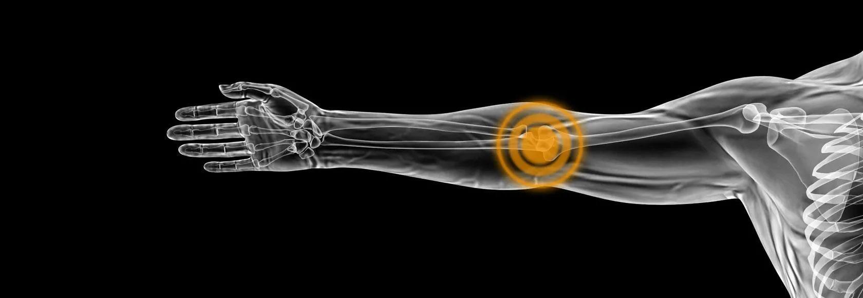
The difference is which side of the elbow hurts. Tennis elbow affects the outside of the elbow, where the tendons that straighten the wrist and fingers attach. Golfer’s elbow affects the inside, where the tendons that bend the wrist attach. Despite the names, most people who get either condition have never played the sport — both are caused by repetitive gripping and forearm use, at work as often as in sport.
The short answer
Tennis elbow
Golfer’s elbow
Medical name
Lateral epicondylitis, or lateral elbow tendinopathy
Medial epicondylitis, or medial elbow tendinopathy
Where it hurts
Outer side of the elbow
Inner side of the elbow
Tendon involved
Common extensor tendon, mainly extensor carpi radialis brevis
Common flexor tendon
Worse when you
Straighten the wrist, grip, lift with palm down
Bend the wrist, grip, twist inwards
Everyday triggers
Carrying shopping, using a screwdriver, typing, lifting a kettle
Swinging a racket or club, throwing, heavy lifting, manual work
How common
The more common of the two
A minority of elbow tendon cases
The two conditions are mirror images across the elbow.
Tennis elbow
Several forearm muscles that straighten the wrist and fingers share a single attachment point on the bony bump on the outside of the elbow. That shared attachment is the common extensor tendon, and one of its components — extensor carpi radialis brevis — is the tendon most often involved.
Despite the name ending in -itis, this is not primarily inflammation. Tissue studies show a degenerative change: the orderly collagen fibres become disorganised, small blood vessels grow into the tendon, and its structure weakens. This matters practically, because it explains why anti-inflammatory tablets often give only limited relief and why loading the tendon gradually works better than resting it completely.
Typical symptoms: pain and tenderness on the outer elbow, often at a very specific point; pain when gripping, shaking hands, turning a door handle or lifting a cup; weakened grip; discomfort spreading down the forearm; and a tendon that is worst first thing or after a period of rest.
Golfer’s elbow
The mirror image. The muscles that bend the wrist and turn the palm downwards attach to the bony bump on the inner elbow, via the common flexor tendon, and the same degenerative process affects them.
Typical symptoms: pain and tenderness on the inner elbow; pain when gripping, bending the wrist or twisting the forearm inwards; and weakened grip. It is less common than tennis elbow — published series put it at around one in five of all epicondylitis cases.
One extra consideration on the inner side: the ulnar nerve passes through a groove immediately behind the medial epicondyle. Pins and needles or numbness in the little and ring fingers points towards nerve irritation rather than, or alongside, tendon problems. That changes what the scan needs to look at.
What ultrasound shows
Both conditions can usually be diagnosed from history and examination. Ultrasound is useful when the diagnosis is uncertain, when symptoms have not settled with initial treatment, when a tear is suspected, or when an injection is planned.
An elbow ultrasound assesses the tendon directly and in real time. A healthy tendon has a neat, striped fibrillar appearance. A tendon affected by tendinopathy typically shows:
Thickening compared with the other side
Darker, less organised areas where the normal fibre pattern has broken down
New blood vessels growing into the tendon, visible with Doppler — a marker of active tendinopathy
Calcium deposits within the tendon or at its attachment
A partial tear — a defined gap in the fibres, which is a different problem from degeneration
Bony changes at the epicondyle
Scanning also allows the other side to be compared and lets the sonographer move the joint while watching, which static imaging cannot do. Published comparisons put ultrasound close to MRI for this region, with the caveat that the result depends heavily on the experience of the person holding the probe.
From our practice
From our practice
On scan, the distinction is anatomical and usually clear. Tennis elbow involves the common extensor tendon on the outer elbow; golfer’s elbow involves the common flexor tendon on the inner side. We scan both sides of the elbow rather than only the side that hurts.
Most of the time, ultrasound confirms what the patient already suspected — they had worked out from where it hurts and what aggravates it, and the scan agrees. But not always. Sometimes it shows a different tendon pattern from the one expected, and sometimes it shows an associated tear rather than straightforward degeneration.
That second finding is the one that changes things. A tear is managed differently from a tendon that is simply degenerate, and it is not something examination reliably distinguishes. It is the main reason a scan earns its place when symptoms are not settling as expected.
Describes a general pattern across the patients we scan, not any individual case.
Treatment
Both conditions are managed the same way, and both usually improve — though slowly. Recovery is measured in months rather than weeks, and that expectation is worth setting early.
First steps. Modify the activities that provoke it rather than stopping using the arm altogether. Complete rest tends to weaken the tendon further. Ice can help after activity. Simple pain relief is reasonable short-term.
Loading exercises. This is the treatment with the best evidence. Progressive, controlled strengthening — often eccentric or isometric exercises — stimulates the tendon to remodel. A physiotherapist will set the programme and progress it. Consistency over several months matters more than intensity.
Bracing. A counterforce strap worn just below the elbow can reduce load on the tendon attachment. It helps some people during aggravating activities; it is not a treatment on its own.
Injections. An ultrasound-guided corticosteroid injection can settle pain in the short term, and guidance makes placement more accurate. The evidence is nuanced: pain relief in the first weeks is often good, but outcomes at a year can be no better — and in some studies worse — than exercise alone. It is best considered when pain is preventing you from doing the rehabilitation, rather than as a substitute for it. Our ultrasound-guided injections start from £250, and we explain why image guidance improves placement accuracy in ultrasound-guided injections: why guidance matters.
Other options. Shockwave therapy and platelet-rich plasma are used for stubborn cases with mixed evidence. Surgery is reserved for a small minority whose symptoms persist beyond six to twelve months of proper conservative treatment.
See a doctor rather than booking a scan if you have: elbow pain following a significant injury or fall; a visibly deformed or badly swollen elbow; a hot, red, swollen joint with fever; numbness or weakness in the hand; or pain that wakes you consistently at night. These point away from simple tendinopathy.
Common questions
How do I tell tennis elbow from golfer’s elbow myself?
Press the bony bumps on each side of your elbow. Tennis elbow is tender on the outside and hurts more when you straighten your wrist against resistance. Golfer’s elbow is tender on the inside and hurts more when you bend your wrist against resistance.
Can I have both at once?
Yes, though it is less common. Some people have tenderness on both sides, particularly with heavy repetitive gripping work.
Do I need a scan to be diagnosed?
Not usually. Most cases are diagnosed clinically. A scan is worth having if the diagnosis is unclear, if symptoms are not improving after a reasonable period of treatment, if a tear is suspected, or if an injection is planned.
How long does it take to get better?
Most people improve over several months with consistent loading exercises. Some cases settle in six to eight weeks; some take a year or more. Slow progress is normal and does not mean the treatment is failing.
Should I stop using my arm completely?
No. Complete rest tends to make tendons weaker. Reduce the specific movements that provoke pain while keeping the arm active, and add progressive loading under guidance.
What does an elbow ultrasound cost and how quickly do I get results?
An elbow ultrasound is £235. You receive instant verbal results from the consultant performing your scan, with the written report following within 24 hours.
CQC-registered clinic · BMUS member · ICO-registered. Reports can be shared securely with your NHS GP or specialist at your request.
References
National Institute for Health and Care Excellence, Clinical Knowledge Summaries. Tennis elbow. Accessed 30 July 2026.
Walz DM et al. Epicondylitis: pathogenesis, imaging, and treatment. RadioGraphics 2010;30(1):167–184.
Sobiech M et al. Ultrasound in the differential diagnosis of medial epicondylalgia and medial elbow pain. Journal of Clinical Medicine 2022. Accessed 30 July 2026.
Coombes BK et al. Efficacy and safety of corticosteroid injections and other injections for management of tendinopathy. The Lancet 2010;376:1751–67.