
Private Breast Screening: Ultrasound vs Mammogram – Which Do You Need?
The short version: your age and your breast density decide. Under 40, dense glandular tissue makes ultrasound the first-line test. Over 40, mammography is the gold standard for screening the whole breast — with ultrasound alongside it to investigate a specific lump or supplement dense tissue. This guide explains why, what each test can and cannot see, and how the NHS screening programme fits in.
First: a new lump is not a screening question
Screening is for people with no symptoms. If you have found a lump, a change in the skin or nipple, or new focal pain, that is an assessment question — UK referral guidance treats an unexplained breast lump as an urgent-referral symptom, and the right first test for a palpable lump is a breast ultrasound, available same-day. Do not wait for a screening invitation to get a symptom checked.
The age-and-density rule, explained
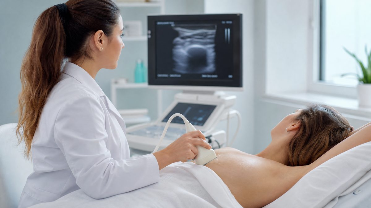
Breast tissue changes with age. Younger breasts are mostly dense, glandular tissue; after around the age of 40 that tissue is gradually replaced by fat. This matters because of how the two tests work. A mammogram is an X-ray: dense tissue and cancers both appear white, so in a young, dense breast a small lump can hide in plain sight. Fatty tissue appears dark, so in an older breast the same lump stands out clearly. Ultrasound works the other way: it images dense tissue well, distinguishes solid lumps from fluid-filled cysts in real time, and checks the lymph nodes under the arm in the same examination — but it is a targeted tool, examining the breast section by section, rather than a standardised whole-breast screening X-ray.
The answer Daniela gives when patients ask "which one do I need?"
It is one of the most common questions in the scanning room, and her standard answer is based on age and tissue density:
"If you are under 40, your breast tissue is typically very dense and glandular. Looking for a small lump on a mammogram in dense tissue is like looking for a snowball in a blizzard. Therefore, ultrasound is our first-line tool. If you are over 40, your tissue naturally becomes fattier, making a mammogram the gold standard for screening the whole breast. However, we still use ultrasound alongside it to investigate a specific lump or if you have very dense breasts."
Shared by Daniela Stan, Consultant Ultrasound Practitioner. General guidance from practice, not individual medical advice — your own history and risk factors can change the right answer, which is exactly what your clinician weighs up.
The two tests, side by side
Breast ultrasound is best for
- Investigating a specific lump, focal pain or nipple change
- Dense breast tissue, at any age
- Telling a fluid-filled cyst from a solid lump, instantly
- Checking the axillary (armpit) lymph nodes
- Women under 40, and during pregnancy or breastfeeding — no radiation
Mammography is best for
- Whole-breast screening of women with no symptoms, especially over 50
- Detecting microcalcifications — tiny calcium specks that can be the earliest sign of change, and which ultrasound generally cannot see
- Standardised, comparable images year on year
- The evidence-backed national screening programme
Neither test "wins" — they answer different questions. That is why women over 40 with a specific lump often sensibly have both: the mammogram for the whole-breast view, the ultrasound to characterise the lump itself. And in genuinely dense breasts, research pooling 29 studies found that adding ultrasound to mammography detects additional cancers the mammogram alone missed — the reason "you have dense breasts" on a screening letter is worth acting on, not filing.
Where the NHS screening programme fits
In the UK, the NHS Breast Screening Programme invites women from age 50 up to their 71st birthday for a mammogram every three years. It is free, evidence-based, and worth attending every time — nothing on this page replaces it. What the programme does not cover is the gaps: women under 50, the three years between invitations, symptoms that appear at any age, and the follow-up of dense-tissue findings. Those gaps are where self-referred ultrasound earns its place — as a complement to the programme, never a substitute for it.
Know your normal — the habit that finds changes early
Whatever your age, the most powerful screening tool you own is familiarity with your own breasts — checking them regularly, at different times of the month, so a genuine change stands out. The changes worth reporting promptly: a new lump or bumpy area in the breast or armpit that feels different from the other side; a change in size, outline or shape; skin dimpling or thickening; nipple discharge that is new for you; a nipple that has become pulled in or points differently; a rash, bleeding or redness. Before menopause, breasts are often naturally tender and lumpy in the days before a period — the signal is change that persists, not lumpiness itself. Our guide to the signs to look for on a self-check covers each one in detail, and if what you have found feels like a cyst or a smooth, mobile lump, the benign breast conditions guide explains what those usually turn out to be.
Family history and ongoing monitoring
A strong family history of breast or ovarian cancer changes the conversation: you may qualify for earlier or more frequent surveillance through NHS family-history and genetics services, and your GP is the route in. Between formal surveillance rounds — or while a benign finding such as a fibroadenoma is being watched — ultrasound is the practical monitoring tool: radiation-free, repeatable, and reported against the previous scan. At Sonoworld the examination includes both breasts and the axillary nodes where indicated, with a BI-RADS-classified report your GP, breast surgeon or family-history clinic can act on directly. For a broader annual check, the Well Woman screening covers the pelvis, upper abdomen, urinary tract and aorta in one visit; it does not include breast ultrasound.
The honest limits, both ways
Ultrasound cannot see the microcalcifications that mammography detects, it depends on the operator examining systematically, and a normal ultrasound in a woman over 50 does not replace her screening mammogram. Mammography, in turn, is less sensitive in dense tissue, involves a small radiation dose, and cannot always say whether a lump is solid or fluid. And neither test diagnoses cancer by itself — imaging finds and describes; only a biopsy answers at cell level. Any clinic that presents one test as universally superior is selling, not informing.
Ultrasound vs mammogram: your questions answered
Which is better — a breast ultrasound or a mammogram?
Neither, universally: they answer different questions. Ultrasound is first-line under 40 and for investigating a specific lump at any age; mammography is the gold standard for whole-breast screening from your 40s onwards. Over 40 with a lump, the two are often used together deliberately.
Can a breast ultrasound replace a mammogram?
No. Ultrasound cannot see microcalcifications — often the earliest mammographic sign of change — and the evidence base for population screening belongs to mammography. Ultrasound complements it: for dense tissue, for lump assessment, and for the years and gaps the programme doesn't cover. Keep every screening invitation.
At what age does NHS breast screening start?
The NHS Breast Screening Programme invites women from age 50 up to their 71st birthday, every three years, for a mammogram. If you are registered with a GP you are invited automatically; after 71 you can still self-refer into the programme.
I'm under 40 and found a lump — which test do I need?
Ultrasound, and promptly. Under 40 your tissue is typically dense, which limits mammography, and UK guidance treats an unexplained lump as an urgent-referral symptom regardless of age. A same-day breast ultrasound characterises the lump — most turn out to be cysts, fibroadenomas or normal glandular tissue — and the report gives your GP exactly what the next step needs.
What if I've been told I have dense breasts?
Take it seriously but calmly: dense tissue is common and normal, but it lowers mammographic sensitivity. Research pooling 29 studies shows that adding ultrasound to mammography in dense breasts finds additional cancers. Practically: keep your mammogram appointments, and discuss supplemental ultrasound with your clinician — or self-refer for one between rounds.
Does Sonoworld do mammograms?
No — we are a specialist ultrasound clinic, and we say so plainly. We provide the ultrasound side: same-day breast ultrasound (£235 one side, £350 both) with lymph-node assessment and a BI-RADS report within 24 hours. If your situation calls for a mammogram, the report says so and your GP or the NHS screening programme is the route — we will never sell you the wrong test.
Need the ultrasound side sorted?
Same-day breast ultrasound with axillary lymph-node assessment, performed personally by Daniela and explained in the room.
Book a breast ultrasound
Well Woman screening (£500)
Self-referral at 29 Weymouth Street, Marylebone · £235 unilateral / £350 bilateral · instant verbal results, BI-RADS written report within 24 hours · 020 3633 4902
Sources and further reading
- NHS Breast Screening Programme — invitation ages and three-year interval; breast awareness guidance. NHS.uk, accessed July 2026.
- NICE Guideline NG12 — Suspected cancer: recognition and referral (2015, amended 15 April 2026): urgent-referral criteria for unexplained breast lumps. Accessed 11 July 2026.
- Rebolj M et al. — Addition of ultrasound to mammography in dense breast tissue: systematic review and meta-analysis of 29 studies. British Journal of Cancer (2018). As cited on our breast ultrasound service page; verified 11 July 2026.
This article is general health information, not a diagnosis or individual medical advice. It was prepared by the Sonoworld clinical team from the practice experience of Daniela Stan, Consultant Ultrasound Practitioner, and against the guidance listed above. A new breast symptom should be assessed promptly rather than waiting for screening. Sonoworld Diagnostic Services, 29 Weymouth Street, Marylebone, London W1G 7DB — CQC-registered.


