Book Scan
MSK · Knee Scan

Private Knee Ultrasound
Scan in London

Expert knee ultrasound imaging for patellar tendinopathy, Baker's cyst, collateral ligament injuries, and periarticular soft tissue pathology. CQC-registered Harley Street clinic, same-day appointments, no GP referral required.

CQC Registered Good No GP Referral Needed Same-Day Results Harley Street, London

Instant verbal results. Written report within 24 hours.

Knee Ultrasound Scan at Sonoworld London
£235All-inclusive price
30mScan duration
24hWritten report
4.9★Patient rating
About This Scan

Expert Musculoskeletal Ultrasound

Musculoskeletal (MSK) ultrasound provides real-time, high-resolution imaging of joints, tendons, ligaments, and soft tissue. Unlike MRI, it allows dynamic assessment — structures can be examined in motion, both sides compared simultaneously, and the sonographer can provide immediate verbal feedback during the examination.

At Sonoworld, all MSK scans are performed by HCPC-registered consultant sonographers with specialist musculoskeletal expertise, using high-resolution GE ultrasound equipment. No GP referral is required, and a comprehensive written report is delivered within 24 hours.

Why patients choose Sonoworld
  • CQC-registered clinic — inspected and rated Good.
  • HCPC-registered consultant sonographers with specialist MSK expertise.
  • Dynamic real-time imaging — structures assessed in motion.
  • Instant verbal results and a written report within 24 hours.
  • No GP referral required — self-refer online or by phone.
  • Transparent all-inclusive pricing at £235.
What We Assess

What We Check During Your Scan

Our consultant sonographers perform a systematic assessment of all relevant structures. Here is what is included in your examination.

Patellar & Quadriceps Tendons

Tendinopathy, partial tears & enthesopathy

Assessment of the patellar tendon and quadriceps tendon for tendinopathy, partial tears, and enthesopathy at the tibial tuberosity and patella. Commonly affected in runners and jumping athletes.

Collateral Ligaments & Soft Tissue

MCL, LCL & periarticular structures

Evaluation of the medial and lateral collateral ligaments for sprains and tears. The periarticular soft tissues including the iliotibial band, pes anserine bursa, and prepatellar bursa are also assessed.

Baker's Cyst & Joint Effusion

Popliteal fossa & posterior knee

Assessment of the popliteal fossa for Baker's cyst (popliteal cyst), joint effusion, and posterior knee soft tissue masses. Baker's cysts are a common cause of posterior knee pain and swelling.

Preparation

How to Prepare for Your Scan

No preparation is required for a knee ultrasound scan. Wear or bring shorts or loose trousers that can be rolled above the knee. You may be asked to bend and extend the knee during the scan for dynamic assessment.

On the day of your scan
  • Arrive 5 minutes before your appointment time.
  • Bring any previous scan reports, X-rays, or MRI results if available.
  • Wear comfortable, loose clothing for the area being scanned.
  • No fasting or special preparation is required.
  • You may bring a companion to your appointment.
The Process

What Happens at Your MSK Scan

1Arrival & Registration

Arrive at our Harley Street clinic. Our team will take a brief clinical history, including your symptoms, duration, and any previous investigations or treatment.

2Dynamic Ultrasound Scan

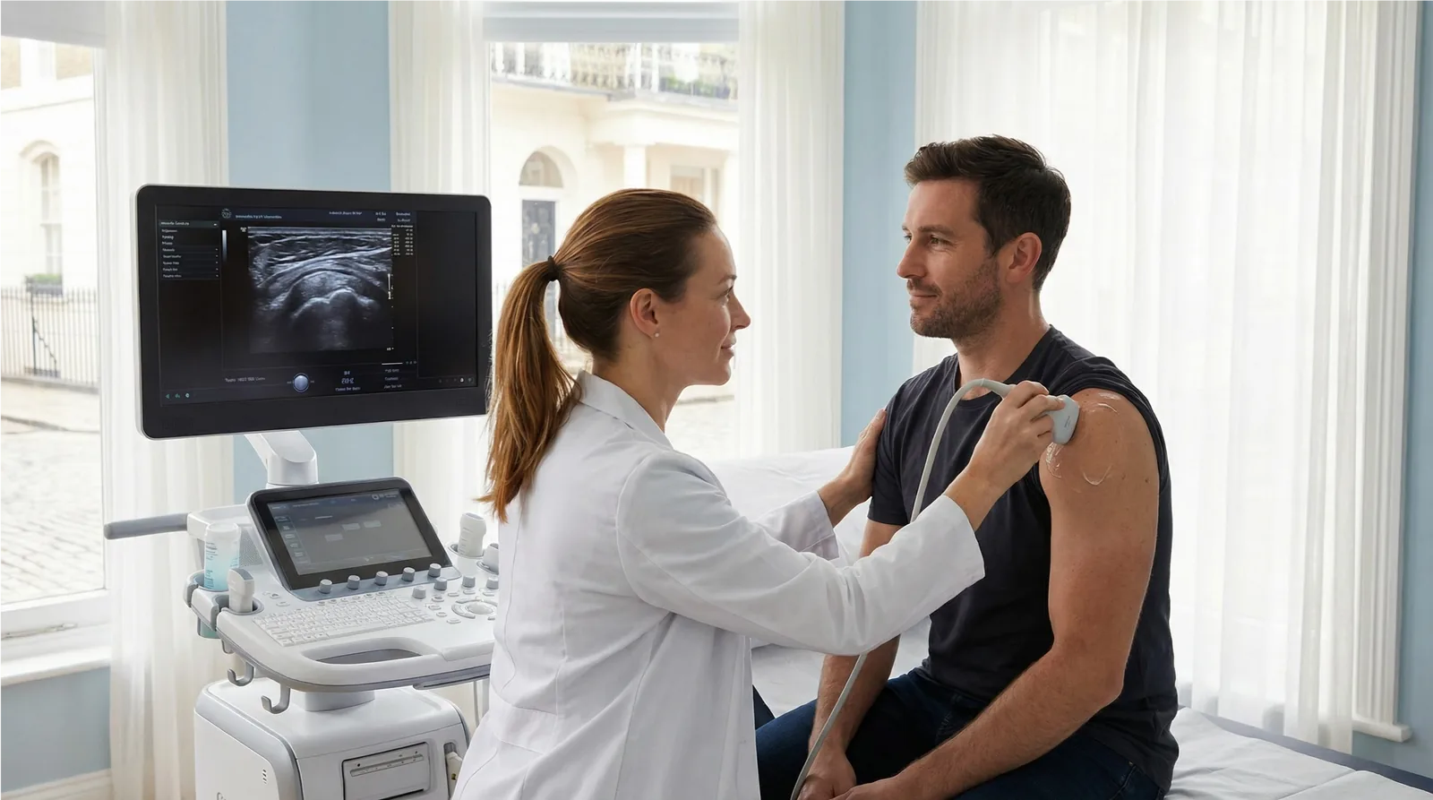
Gel is applied to the skin and your consultant sonographer scans the affected area using a high-resolution probe. You may be asked to move the joint during the scan for dynamic assessment.

3Immediate Verbal Feedback

Your sonographer explains their findings immediately after the scan, answering any questions about what was seen and what it means for your management.

4Written Report Within 24 Hours

A comprehensive written report is emailed to you within 24 hours. You may share this directly with your GP, physiotherapist, or specialist.

Pricing

Scan Pricing

£235All-inclusive — no hidden fees
  • Consultant sonographer examination
  • Dynamic real-time ultrasound imaging
  • Immediate verbal results
  • Comprehensive written report within 24 hours
  • Report shared with your GP or specialist on request
Book Your Scan

View all MSK scan types on the MSK Ultrasound Scans Hub.

FAQs

Frequently Asked Questions

What does a knee ultrasound scan show?

A knee ultrasound scan provides detailed imaging of the patellar tendon, quadriceps tendon, collateral ligaments, Baker's cyst, bursae, and periarticular soft tissues. It does not assess the menisci or cruciate ligaments — MRI is required for these structures.

Can ultrasound detect a meniscal tear?

Ultrasound has limited ability to assess the menisci, as they are partially obscured by bone. MRI is the preferred modality for meniscal assessment. However, ultrasound is excellent for assessing tendons, ligaments, bursae, and Baker's cysts.

Do I need a GP referral for a knee scan?

No. At Sonoworld, you can self-refer directly without a GP referral. Simply book online or call our team.

How long does a knee ultrasound scan take?

The scan typically takes 20–30 minutes. Your sonographer will provide immediate verbal feedback, and a written report will be emailed within 24 hours.

What is a Baker's cyst and can ultrasound diagnose it?

A Baker's cyst is a fluid-filled sac at the back of the knee, often associated with joint effusion or meniscal pathology. Ultrasound is the gold-standard investigation and can confirm the diagnosis, measure the cyst, and assess for complications such as rupture.

Book Your Knee Ultrasound Scan — £235 All-Inclusive
crossmenu
Not sure which scan you need?
Sono AISonoworld Scan Advisor
Tap to Call

If imaging confirms inflammation or soft-tissue injury, Sonoworld also offers ultrasound-guided cortisone injection for knee pain — a precise, same-day treatment performed under real-time ultrasound guidance.

If your knee scan reveals inflammation or a painful soft tissue condition, you may benefit from an ultrasound-guided cortisone injection for knee pain at Sonoworld. Our precision-guided injections are performed under real-time ultrasound imaging for accurate placement and faster relief.