Breast Cancer Symptoms: 12 Signs to Know
Breast cancer does not always present as a lump. A consultant sonographer explains the 12 signs that warrant a conversation with your GP, and what a private breast ultrasound adds to the assessment.
- Women's Health
- Breast Health
- Updated July 2026
Why Awareness of Symptoms Matters
Breast cancer is the most common cancer among women in the UK — approximately 1 in 7 women will develop it in their lifetime. Early detection significantly improves treatment outcomes: the five-year survival rate for stage 1 breast cancer is above 98%, compared to approximately 26% for stage 4. The gap between those two numbers is largely explained by how early the cancer was found.
The NHS breast screening programme invites women aged 50–71 for mammography every three years. That leaves a significant window — and a significant proportion of women — outside routine screening. Understanding what to look for, and acting on it promptly, is what closes that gap.
This guide covers the 12 symptoms that should prompt a GP appointment, the role of breast ultrasound in the assessment, and when to seek urgent review rather than a routine appointment.
The 12 Breast Cancer Symptoms to Know
Not all of these symptoms indicate cancer — most do not. But all of them, when new, persistent, or changing, warrant medical assessment. Do not wait for pain: most early breast cancers are painless.
1. A New Lump or Thickening
The most commonly recognised symptom. A new lump in the breast or armpit — particularly one that is hard, has irregular edges, and does not move easily — should be assessed promptly. Not all lumps are cancerous, but all new lumps deserve investigation.
2. Dimpling or Puckering of the Skin
Changes to the skin surface — dimpling, puckering, or a texture resembling orange peel (peau d'orange) — can indicate an underlying mass pulling on the skin's ligaments. This is a red flag that should not be attributed to weight change or ageing without investigation.
3. Change in Breast Size or Shape
Any unexplained, asymmetrical change in the size or shape of one breast — particularly if it has appeared over weeks rather than months — warrants assessment. Gradual changes over years are usually benign; rapid or asymmetric changes are not.
4. Nipple Discharge
Spontaneous nipple discharge — particularly if it is blood-stained, clear, or occurs from one breast only — requires evaluation. Discharge that only occurs when the nipple is squeezed is less concerning but should still be mentioned to your GP if it is new.
5. Nipple Inversion or Change in Direction
A nipple that suddenly turns inwards (inverts), changes its direction, or develops a persistent rash or scaling may indicate an underlying structural change. A nipple that has always been inverted is not a concern; a nipple that has recently changed is.
6. Rash on the Nipple or Areola
A persistent rash, redness, or scaling on the nipple or areola that does not respond to topical treatment should be assessed. Paget's disease of the nipple — a rare form of breast cancer — can present exactly like eczema.
7. Redness or Warmth of the Breast
Unexplained redness, warmth, or swelling of the breast — particularly if it is not associated with an infection — can be a symptom of inflammatory breast cancer, a rare but aggressive form of the disease. This should be assessed urgently, not monitored at home.
8. Persistent Itchiness
Persistent, unexplained itching of the breast or nipple area — particularly if accompanied by any of the other symptoms on this list — should be discussed with a GP. On its own, itchiness is rarely a sign of cancer; in combination with other changes, it is worth investigating.
9. Breast or Armpit Pain
Cyclical breast pain (pain that tracks with the menstrual cycle) is common and usually hormonal. Non-cyclical, localised pain — particularly if it is persistent, worsening, or accompanied by a tender area on examination — deserves investigation. Pain alone is rarely the first sign of breast cancer, but it is not a reason to dismiss other symptoms.
10. Swelling in the Armpit or Collarbone Area
Swollen lymph nodes in the armpit or around the collarbone can indicate that breast cancer has spread to the lymphatic system. A new, persistent lump in the armpit — even without any breast symptoms — should be assessed.
11. Thickening of Breast Tissue
A noticeable thickening or hardening of part of the breast tissue — an area that feels different from the surrounding tissue and has not been there before — is worth investigating, even if it does not feel like a discrete lump.
12. Any Persistent Change That Is Unusual for You
This is the most important item on the list. You know your breasts. Any persistent change — even one that does not fit neatly into the categories above — that is new and has not resolved after two to three weeks warrants a GP appointment. Trust your instincts.
When to Seek Urgent Review
NICE guideline NG12 sets a two-week wait standard for suspected breast cancer. Your GP should refer you urgently (within two weeks) if you have:
- A new breast lump in a woman aged 30 or over
- Unilateral nipple discharge in a woman aged 50 or over
- Skin changes suggestive of breast cancer (dimpling, peau d'orange, nipple changes)
- An unexplained lump in the axilla in a woman aged 30 or over
If you are under 30 with a new breast lump, a GP appointment is still appropriate — the two-week wait criteria are different, but the lump should still be assessed.
A private breast ultrasound at Sonoworld can be booked the same day, without a GP referral, and provides a same-day structural assessment. It does not replace a GP appointment or a two-week wait referral — but it provides information that can support and accelerate the clinical pathway.
The Role of Breast Ultrasound in Symptom Assessment
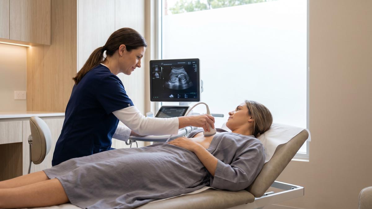
Breast ultrasound is the first-line imaging modality for any palpable breast lump, focal pain, or nipple discharge in women under 40, and is used alongside mammography in older women when a specific area needs further characterisation. It does not use ionising radiation, is safe at any age and in pregnancy, and provides real-time images that can be explained verbally during the scan.
At Sonoworld, findings are reported using the ACR BI-RADS lexicon — a standardised classification from 1 (negative) to 5 (highly suspicious) that makes the report directly actionable for any onward referral. The written report is sent the same day. For a full description of what the scan involves, see our guide: Private Breast Ultrasound Scan — What to Expect.
For a comparison of breast ultrasound and mammography, see: Breast Ultrasound vs Mammogram: Which Do You Need?
From Our Practice
The most common presentation in our breast-imaging casebook is a woman who has found something and wants an answer the same day. The "something" is often a lump — but a significant proportion of self-referrals are for symptoms that do not fit the classic lump pattern: a persistent area of tenderness, a subtle skin change, a nipple that has shifted slightly, or simply a feeling that something is not right.
The pattern we observe most consistently is that women who self-refer with non-lump symptoms have often already been to their GP and been reassured. They book a private scan because the reassurance did not settle the concern — and in a meaningful proportion of these cases, the scan finds something that the clinical examination did not. Not always cancer; often a cyst, a fibroadenoma, or an area of fibrocystic change that explains the symptom. But the finding is real, and the patient leaves with an answer rather than a continued uncertainty.
The second pattern is women in their 30s who have dense breast tissue and have been told that mammography is not the right first test for their age. Ultrasound is the appropriate modality in this group, and it is the one we use. Dense breast tissue is not an obstacle for ultrasound — it is the condition in which ultrasound performs best.
Observations above are drawn from composite patterns across our patient population, not from any individual case. They are not a substitute for individual medical advice or a GP assessment.
How to Check Your Breasts
There is no single correct technique for breast self-examination — the most important thing is to check regularly and to know what is normal for you. The NHS recommends checking your breasts at least once a month, at any time that is convenient. The key steps are:
- Look in the mirror with arms at your sides, then raised above your head. Look for changes in size, shape, or skin texture, and check the nipples for any changes in direction or appearance.
- Feel your breasts using the pads of your fingers in a circular motion, covering the entire breast area from the collarbone to the abdomen and into the armpit. Use a firm, smooth touch.
- Check both breasts and both armpits. A lump in the armpit is as significant as a lump in the breast.
- Report any new or persistent change to your GP promptly. Do not wait to see if it resolves.
Sources
- National Institute for Health and Care Excellence. Suspected cancer: recognition and referral (NG12). NICE, 2015 (updated 2023). nice.org.uk/guidance/ng12
- NHS. Symptoms of breast cancer in women. Accessed July 2026. nhs.uk
- Cancer Research UK. Symptoms of breast cancer. Accessed July 2026. cancerresearchuk.org
- Sonoworld clinical imaging team. Composite practice patterns — Sonoworld Diagnostic Services, Marylebone, London. July 2026.
Book a Breast Ultrasound Scan
£235 (one breast) · £350 (both breasts). Same-day appointments available. No GP referral needed. BI-RADS report sent the same day.
29 Weymouth Street, Marylebone, London W1G 7DB | 020 3633 4902