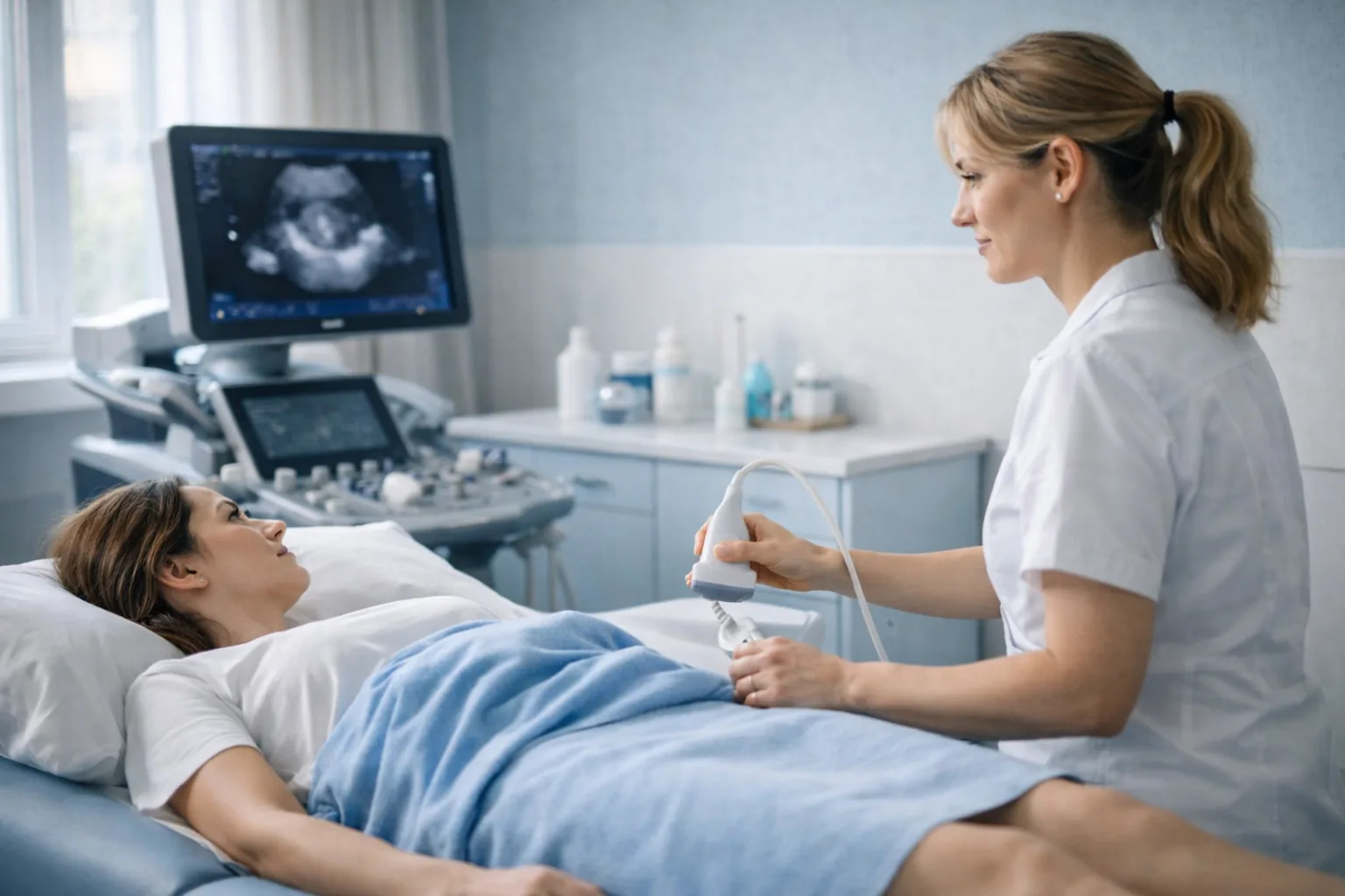
A pelvic and transvaginal ultrasound performed to a dedicated endometriosis protocol: the ovaries for endometriomas, the uterus for adenomyosis, and the space behind the uterus for deep endometriosis and adhesions. Performed by a female consultant ultrasound practitioner at our CQC-registered Marylebone clinic. Same-day appointments, no GP referral required.
CQC Registered Good No GP Referral Needed Female Consultant Practitioner Marylebone, London
Booked as a pelvic ultrasound appointment. Instant verbal results. Written report within 24 hours.
£235All-inclusive price
20–30mScan duration
24hWritten report
CQCRated Good
About This Scan
A Pelvic Scan Built Around Endometriosis
Endometriosis affects around one in ten women of reproductive age, and many wait years for a diagnosis because cyclical pelvic pain, painful periods and pain during sex are so easily put down to other causes. NICE guidance (NG73) names transvaginal ultrasound as the first investigation to offer when endometriosis is suspected, even when a physical examination is normal.
A standard pelvic scan looks at the uterus and ovaries. An endometriosis scan goes further. It uses the transvaginal probe to examine the posterior compartment behind the uterus, tests whether the pelvic organs slide freely over each other (the sliding sign), and looks specifically for the three findings that matter most: ovarian endometriomas, adenomyosis within the uterine wall, and deep endometriosis nodules on the uterosacral ligaments, rectovaginal septum, bowel or bladder.
One honest limit: ultrasound cannot see superficial endometriosis on the peritoneal lining. A normal scan therefore lowers the likelihood of significant disease but does not rule endometriosis out, and laparoscopy remains the definitive test. Your report says clearly what was and was not seen so that your GP or gynaecologist can decide the next step.
Why patients choose Sonoworld
CQC-registered clinic — inspected and rated Good.
Female consultant ultrasound practitioner with a gynaecological imaging focus.
Dedicated protocol: posterior compartment and sliding sign, not just uterus and ovaries.
Instant verbal results and a written report within 24 hours.
No GP referral required — self-refer online or by phone.
Transparent all-inclusive pricing at £235.
Who It Is For
When to Book an Endometriosis Scan
This scan suits anyone with symptoms that follow the menstrual cycle, anyone whose standard pelvic scan was normal but whose pain continues, and anyone who wants a structured assessment before a gynaecology appointment.
Common reasons to book
Pelvic pain that comes and goes with your cycle, or has lasted six months or more.
Periods painful enough to disrupt work, sleep or exercise.
Deep pain during or after sex.
Painful bowel movements, bloating, rectal bleeding or bladder urgency around your period.
Difficulty conceiving, with or without pain.
A known endometrioma or adenomyosis that needs monitoring.
A family history of endometriosis in a mother or sister.
What We Assess
What We Check During Your Scan
The examination follows a systematic protocol so that every compartment of the pelvis is documented, not only the areas that happen to look abnormal.
Ovaries
Endometriomas (“chocolate cysts”)
Cysts filled with old blood have a characteristic ground-glass appearance. We measure them, describe their features and note whether they sit on one or both sides, which influences the likelihood of deeper disease.
Uterus
Adenomyosis
Endometrial tissue within the muscle wall produces an enlarged, asymmetric uterus with small cysts in the wall and a blurred junctional zone. It often coexists with endometriosis and explains heavy, painful periods.
Posterior Compartment
Deep endometriosis and the sliding sign
The uterosacral ligaments, rectovaginal septum, bowel wall and pouch of Douglas are examined for nodules. Gentle probe pressure shows whether the uterus and bowel slide freely; a negative sliding sign points to adhesions.
Everything Else
Fibroids, polyps and other cysts
Conditions that mimic or accompany endometriosis are documented in the same visit, so the report gives your clinician a complete picture of the pelvis rather than a single answer.
Preparation
How to Prepare for Your Scan
Drink about a litre of water an hour before your appointment and do not empty your bladder: the first part of the scan is performed through the abdomen and needs a full bladder. You will be asked to empty it before the transvaginal part. The scan can be done at any point in your cycle, and you should continue any medication, including hormonal treatment, as normal.
On the day of your scan
Arrive 5 minutes before your appointment time.
Bring previous scan reports, gynaecology letters or a symptom diary if you keep one.
Wear loose clothing that gives easy access to the lower abdomen.
No fasting is needed.
The transvaginal part is explained first and performed only with your consent; you can decline it or stop at any time.
You may bring a companion or chaperone.
The Process
What Happens at Your Endometriosis Scan
1Clinical History
Your practitioner asks about the pattern, timing and severity of your symptoms, previous investigations and any fertility plans. This shapes the examination.
2Transabdominal Scan
With a full bladder, the uterus, ovaries and bladder wall are assessed from the outside to give an overview of the pelvis.
3Transvaginal Scan
With your consent, a slim internal probe gives high-resolution images of the uterine wall, endometrium and ovaries, and detects endometriomas and adenomyosis that a transabdominal scan can miss.
4Sliding Sign and Posterior Compartment
The uterosacral ligaments, rectovaginal septum and pouch of Douglas are examined systematically and the sliding sign is tested for adhesions.
5Results and Written Report
Findings are explained straight away. A written report with images follows within 24 hours, structured for your GP, gynaecologist or a specialist endometriosis centre.
Pricing
Scan Pricing
£235All-inclusive — no hidden fees
Consultant ultrasound practitioner examination
Transabdominal and transvaginal assessment with the endometriosis protocol
Immediate verbal results
Written report with images within 24 hours
Report shared with your GP or specialist on request
The scan is booked as a pelvic ultrasound appointment; tell us at booking or on arrival that you would like the endometriosis protocol. Compare all women’s health examinations on the Women’s Health Scans Hub.
FAQs
Frequently Asked Questions
Can an ultrasound scan diagnose endometriosis?
It can identify the forms that matter most: endometriomas on the ovaries, adenomyosis in the uterine wall, and deep endometriosis nodules behind the uterus. It cannot see superficial endometriosis on the peritoneum, so a normal scan does not rule the condition out. Laparoscopy remains the definitive test when symptoms persist.
How is this different from a standard pelvic scan?
A standard pelvic scan assesses the uterus and ovaries. The endometriosis protocol adds a systematic examination of the posterior compartment, the sliding sign test for adhesions and specific assessment for adenomyosis and deep nodules. It takes a little longer and the report is structured for endometriosis referral pathways.
Is the transvaginal part necessary?
It is strongly recommended because the internal probe sits much closer to the ovaries and the space behind the uterus and sees far more detail. It is always explained first and performed only with your consent. If you prefer a transabdominal-only scan we will do that, with the limits noted in your report.
When in my cycle should I have the scan?
Any time. Some women prefer to avoid the heaviest days of their period for comfort, but the findings that matter do not depend on cycle day.
What happens if the scan is normal but I still have symptoms?
Your report will say so clearly. The next step is usually a gynaecology referral, which your GP can make with the report, or a referral to a specialist endometriosis centre where laparoscopy can be considered.
Do I need a GP referral?
No. You can self-refer and book online or by phone. The report can be shared with your GP or specialist afterwards.