Operational rationale
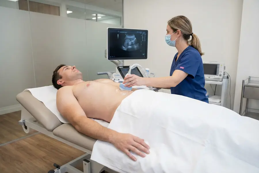
Standing, supine and Valsalva on every scan
The most common reason a hernia is missed on imaging is a static scan performed only with the patient lying down. A small inguinal or femoral hernia can sit completely reduced inside the canal at rest and only become visible when intra-abdominal pressure rises. The published protocol guidance from the American Journal of Roentgenology is explicit: groin hernias must be assessed during Valsalva and documented in two orthogonal planes to avoid diagnostic pitfalls.2
We adopt this as a non-negotiable in our hernia scan protocol. Every patient is scanned at rest, then with a cough, then with a sustained Valsalva, and then a second time standing up. This adds about five minutes to the scan and occasionally turns a negative supine examination into a clearly demonstrated direct inguinal hernia. We would rather take the extra time than send a patient home with a falsely reassuring report.
Clinical pattern
The contralateral groin almost always gets imaged
When patients present with a unilateral groin lump, it is tempting to scan only the symptomatic side. We do not. A cross-sectional study of asymptomatic men aged 45–67 found that 16% had an unsuspected inguinal hernia on ultrasound, with 4% bilateral.4 A meaningful minority of our patients arrive convinced they have a one-sided problem and leave with a clearer picture: an obvious symptomatic hernia on one side, and a small early defect on the other that the surgeon can address at the same operation. Our bilateral scan price (£350) reflects this; it is rarely a wasted second image.
Reasoning
Why we keep the verbal result and the surgical report separate
At the end of every scan, our sonographer turns the screen, walks you through what you can see and what it means, and answers your questions. That conversation is not the diagnosis: it is reassurance and orientation. The diagnosis is the written report, prepared and signed off the same day or next morning, and structured so a hernia surgeon can act on it without re-imaging: type, side, size of defect, contents, reducibility, relationship to the inferior epigastric vessels for inguinal hernias. Most of our patients hand the report directly to the surgeon they have already chosen. A small number ask us for an onward referral; we keep a list of London hernia surgeons we have worked with and will write directly with consent.
Patterns observed across our hernia scan caseload, not individual patient histories. Anonymised composite scenarios; no patient-identifying detail.