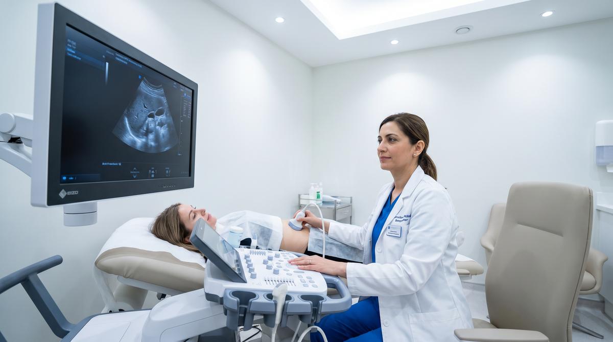
What Is an Ultrasound Scan?
A consultant sonographer explains how ultrasound works, what it can detect, whether it is safe, and when a private scan is worth considering — without the NHS wait.
- No ionising radiation
- Same-day results
- CQC registered clinic
- Consultant-led
Or call us: 020 3633 4902 — same-day appointments available
What Is an Ultrasound Scan?
An ultrasound scan — also called sonography or diagnostic medical sonography — is a medical imaging technique that uses high-frequency sound waves to create real-time images of the inside of the body. Unlike X-rays or CT scans, ultrasound uses no ionising radiation whatsoever, making it one of the safest diagnostic tools available in modern medicine.
The technique was first developed for clinical use in the 1950s and has since become one of the most widely used diagnostic methods globally. It is the first-line imaging choice for assessing soft tissues, organs, blood vessels, and developing pregnancies — precisely because it is safe, painless, and provides immediate results.
At Sonoworld, every scan is performed by a consultant-level practitioner using high-resolution equipment. You receive verbal results during your appointment and a written report within 24 hours. For a full overview of what we scan, see our appointments page.
Quick Reference
- Technology: High-frequency sound waves (2–18 MHz)
- Radiation: None — completely safe for all ages
- Duration: 20–45 minutes depending on scan type
- Results: Verbal during appointment; written within 24 hours
- Preparation: Varies by scan type — see our preparation guide
How Does Ultrasound Work?
The physics behind ultrasound is straightforward. A small handheld device called a transducer — or probe — emits pulses of high-frequency sound waves into the body. These sound waves travel through soft tissues and fluids, and when they encounter a boundary between different tissue types (for example, between the liver and a cyst), they reflect back to the transducer as echoes.
A computer processes the timing and intensity of these returning echoes and converts them into a real-time image on the screen. Because different tissues reflect sound differently, the resulting image distinguishes between solid structures, fluid-filled cavities, and blood flow — all without any radiation.
- Gel is applied to the skin. A water-based gel eliminates air pockets between the probe and your skin, which would otherwise block the sound waves.
- The transducer emits sound waves. The probe is moved gently over the skin. It emits pulses of sound at frequencies between 2 and 18 MHz — far above the range of human hearing.
- Echoes return to the probe. Sound waves bounce back from tissue boundaries. The probe detects these returning echoes and sends the data to the ultrasound machine.
- Real-time images are generated. The machine converts the echo data into a live image. Your practitioner interprets this in real time and explains what they are seeing during the scan.
- You receive your results. Your practitioner provides verbal findings immediately. A detailed written report follows within 24 hours.
Types of Ultrasound Used at Sonoworld
Different scan types use the same underlying technology but vary in how the probe is positioned and what it measures.
- B-mode (2D): The standard greyscale image most people recognise — used for abdominal, pelvic, and musculoskeletal assessments.
- Doppler: Measures blood flow direction and velocity — essential for vascular assessments such as DVT scans and carotid artery scans.
- Transvaginal (TV): An internal probe provides higher-resolution images of the uterus and ovaries — used for pelvic scans and fertility assessments.
- Echocardiography: Specialist cardiac ultrasound that assesses heart structure and function — see our echocardiogram page.
- Musculoskeletal (MSK): High-frequency probes assess tendons, ligaments, and muscles — used for MSK scans and ultrasound-guided injections.
Is Ultrasound Safe?
Yes — diagnostic ultrasound has an excellent safety record spanning more than 60 years of clinical use. The British Medical Ultrasound Society (BMUS) states that there is no confirmed evidence that diagnostic ultrasound has caused harm to any patient, including during pregnancy. This conclusion is supported by extensive laboratory and epidemiological research. [Verified: BMUS Guidelines for the Safe Use of Diagnostic Ultrasound Equipment, accessed July 2026]
Why Ultrasound Is Safe
- No ionising radiation: Sound waves carry no ionising energy and cannot damage DNA or cells.
- Safe in pregnancy: Ultrasound is the standard imaging modality throughout pregnancy and is used routinely by the NHS from the first trimester.
- Safe for all ages: From newborns to elderly patients, there are no age-based contraindications to diagnostic ultrasound.
- Repeatable: Scans can be performed as many times as clinically indicated without any cumulative risk.
- Non-invasive: External ultrasound requires no needles, no contrast agents, and no anaesthetic.
Ultrasound vs Other Imaging
| Modality | Radiation? | Soft Tissue Detail | Same-Day Access |
|---|---|---|---|
| Ultrasound | None | Excellent | Yes |
| X-ray | Yes (low) | Limited | Yes |
| CT scan | Yes (significant) | Good | Possible privately |
| MRI | None | Excellent | Possible privately |
MRI also uses no radiation but is significantly more expensive and less accessible for same-day appointments.
What Can an Ultrasound Scan Detect?
Ultrasound is the first-line imaging investigation for a wide range of conditions across multiple organ systems. It excels at assessing soft tissues, fluid-filled structures, and blood flow — areas where X-rays provide limited information.
Women's Health
- Ovarian cysts and polycystic ovaries
- Uterine fibroids and endometrial thickening
- Fertility assessment and follicle tracking
- Breast lumps and tissue changes
Men's Health
- Testicular lumps and varicoceles
- Prostate and bladder assessment
- Kidney stones and renal tract
- Hernia confirmation and assessment
Abdominal
- Liver, gallbladder, and gallstones
- Pancreas, spleen, and kidneys
- Thyroid and neck lumps
- Abdominal aortic aneurysm (AAA) screening
Vascular & Cardiac
- Deep vein thrombosis (DVT)
- Carotid artery disease and stroke risk
- Heart structure and function (echocardiogram)
- Peripheral arterial and venous disease
MSK
- Rotator cuff and shoulder tendons
- Knee, hip, elbow, and ankle tendons
- Plantar fasciitis and Achilles tendon
- Ultrasound-guided cortisone injections
Pregnancy
- Early pregnancy viability scans
- Dating and nuchal translucency scans
- Growth and wellbeing scans
- 4D bonding scans
From Our Practice
One pattern we see consistently at Sonoworld is patients who arrive having already had an NHS ultrasound — but who were not given a verbal explanation during the scan. They received a letter days later, addressed to their GP, summarising findings they had not yet seen. By the time they reached us, they had spent a week searching the internet for interpretations of terms they did not understand.
The verbal explanation during the scan is not a luxury add-on at a private clinic — it is a clinical communication standard. When a consultant sonographer explains what they are seeing in real time, the patient leaves the appointment with accurate information rather than an anxiety-driven interpretation of a report written for another clinician. In our experience, this single difference — the conversation during the scan — is what patients cite most often when asked why they chose private.
In other cases, the scan identifies something that requires onward referral. Because the patient has a written report in hand, the GP or specialist can act immediately rather than repeating the imaging. The report travels with the patient and shortens the diagnostic pathway.
All observations above are drawn from composite patterns across our patient population, not from any individual case. They are intended to illustrate typical clinical scenarios, not to provide diagnostic guidance. Always seek individual professional assessment for your own symptoms.
Private Ultrasound vs NHS Ultrasound
The clinical quality of the scan itself is the same whether performed privately or on the NHS — both use the same technology and the same clinical standards. The difference lies in access, speed, and the appointment experience.
| NHS Ultrasound | Private Ultrasound at Sonoworld | |
|---|---|---|
| Referral | GP referral required in most cases | No referral required — self-refer directly |
| Waiting time | Typically 6–15 weeks (Jan 2026: 21% waited over 6 weeks — Nuffield Trust) | Same-day and next-day appointments available |
| Results | Sent to your GP; you may wait days for a summary | Verbal results during your appointment |
| Written report | Sent to referring clinician | Sent directly to you within 24 hours |
| Appointment length | Typically 15–20 minutes | Consultant-led, unhurried appointments |
| Cost | Free at point of use | From £235 — see our prices page |
| Insurance | N/A | AXA, Healix, WPA accepted |
When a Private Scan Makes Sense
A private scan is worth considering when you cannot wait for an NHS appointment, when you want to understand a symptom before your GP appointment, when you need a scan not covered by your NHS referral, or when you simply want the reassurance of a same-day result. See our transparent pricing page for all-inclusive fees with no hidden costs.
What to Expect at Your Appointment
Most patients are surprised by how straightforward and comfortable a private ultrasound appointment is. Here is what typically happens from the moment you arrive to when you leave.
- Arrival and check-in. You arrive at our central London clinic. There is no long wait — your appointment is scheduled to start on time.
- Brief clinical history. Your practitioner takes a brief history — why you are having the scan, any relevant symptoms, and any previous imaging. This takes 3–5 minutes and ensures the scan is targeted correctly.
- The scan itself. You lie on the examination couch. Gel is applied to the area being scanned. The probe is moved gently over the skin. Most scans take 20–30 minutes. You can see the screen throughout.
- Verbal results. Your practitioner explains what they found during the scan — in plain language, with time for your questions. You leave the clinic knowing what was seen.
- Written report within 24 hours. A detailed written report is sent to you within 24 hours. This can be shared with your GP, specialist, or insurance provider.
Preparation Varies by Scan Type
Some scans require specific preparation — for example, a full bladder for pelvic scans, or fasting for abdominal scans. Others require no preparation at all. See our full ultrasound preparation guide for scan-specific instructions.
Frequently Asked Questions
Answers to the questions we are most commonly asked about ultrasound scans.
Do I need a GP referral for a private ultrasound scan?
No. At Sonoworld you can self-refer for any scan on our menu — including specialist assessments such as echocardiograms and MSK scans. You do not need to see your GP first. Simply book online or call us on 020 3633 4902.
Is a private ultrasound scan the same quality as an NHS scan?
Yes. The underlying technology and clinical standards are identical. The difference is access speed, the verbal explanation during the scan, and the written report sent directly to you within 24 hours — rather than to your GP days later.
Are private ultrasound scans safe?
Yes. Diagnostic ultrasound uses sound waves, not radiation. The British Medical Ultrasound Society (BMUS) states there is no confirmed evidence that diagnostic ultrasound has caused harm to any patient, including during pregnancy. There is no cumulative dose and no contraindication based on frequency of use. Always seek individual professional assessment for your specific clinical situation.
How long does an ultrasound scan take?
Most scans at Sonoworld take 20–45 minutes, including the clinical history and verbal results. More complex assessments such as echocardiograms or full-body screening packages take longer. Your booking confirmation will include the expected duration for your specific scan.
Will the scan be painful?
External ultrasound is painless. You will feel the probe moving over your skin and the cool gel applied beforehand. Transvaginal ultrasound uses an internal probe and may cause mild discomfort, though it should not be painful. If you experience pain during any scan, tell your practitioner immediately. Always discuss any concerns with your clinician before the scan begins.
Can ultrasound detect cancer?
Ultrasound can identify abnormal tissue that may require further investigation — including suspicious masses in the thyroid, breast, testes, liver, and ovaries. It cannot diagnose cancer definitively; a tissue biopsy is required for a definitive diagnosis. If your scan identifies something concerning, your written report will include a recommendation for the appropriate next step. Always seek individual professional assessment for any suspected abnormality.
How much does a private ultrasound scan cost?
At Sonoworld, private ultrasound scans start from £235. The price includes the consultation, the scan, and a written report — there are no hidden fees. Screening packages start from £500. See our full prices page for a complete breakdown by scan type.
What is the difference between a 2D, 3D, and 4D ultrasound?
2D (B-mode) is the standard greyscale image used for diagnostic assessments. 3D ultrasound reconstructs a static three-dimensional image from multiple 2D slices. 4D adds real-time movement to the 3D image — most commonly used for pregnancy bonding scans. Diagnostic assessments at Sonoworld use 2D and Doppler imaging; our 4D baby scan is available for pregnancy bonding.
Sources and Further Reading
- British Medical Ultrasound Society (BMUS). Guidelines for the Safe Use of Diagnostic Ultrasound Equipment. BMUS Safety Committee. Accessed July 2026. bmus.org
- Nuffield Trust. Diagnostic Test Waiting Times. Published April 2026. Accessed July 2026. nuffieldtrust.org.uk — Source for 21% waiting over 6 weeks for ultrasound, January 2026.
- World Federation for Ultrasound in Medicine and Biology (WFUMB). Clinical Safety Statement for Diagnostic Ultrasound. 2024. Accessed July 2026. wfumb.org
- Society of Radiographers & BMUS. Guidelines for Professional Ultrasound Practice. 2021. Accessed July 2026. bmus.org
- Sonoworld clinical imaging team. Composite practice patterns — Sonoworld Diagnostic Services, Marylebone, London. July 2026.
Clinical Governance
Sonoworld Diagnostic Services is registered with the Care Quality Commission (CQC) and rated Good. All practitioners are registered with the Health and Care Professions Council (HCPC). The clinic is a member of the British Medical Ultrasound Society (BMUS) and registered with the Information Commissioner's Office (ICO).
All health claims on this page comply with the CAP Code. No prescription-only medicines are advertised. Clinical content has been reviewed by the Sonoworld clinical team. Pre-publication sign-off by a named, registered clinician is required before any clinical claim is published — Requires human sign-off.
Your scan report is sent directly to you and is your property. You choose whether to share it with your GP or specialist. Data is handled in accordance with UK GDPR and ICO guidance.
For full governance information, see our About Sonoworld page.
Ready to Book Your Scan?
Same-day and next-day appointments available in central London. No referral needed.
29 Weymouth Street, Marylebone, London W1G 7DB