Cryptogenic Stroke: What Tests Find a Hidden Cause?
By the Sonoworld clinical team, 29 Weymouth Street, Marylebone · CQC-registered clinic · Medically reviewed by Hootan Mohsenian, MSc — Advanced Practitioner in Echocardiography — 31 July 2026 · Last reviewed: 31 July 2026
A cryptogenic stroke is a stroke where the standard investigations have not found a cause. Around one in four ischaemic strokes falls into this group. Because the heart is one of the places a clot can come from, cardiac tests — an echocardiogram, a carotid ultrasound and longer heart rhythm monitoring — often form part of the search. These tests can identify a possible cardiac source. They cannot, on their own, prove that the heart caused the stroke.
Call 999 immediately if you or someone else has new stroke symptoms. Use the FAST test: Face drooping on one side, Arm weakness, Speech difficulty, Time to call 999. This applies even if the symptoms pass quickly — a transient ischaemic attack is a warning that needs urgent assessment, not a private scan.
What a cryptogenic stroke means
An ischaemic stroke happens when the blood supply to part of the brain is blocked, usually by a clot. After a stroke, the hospital team works through a set of investigations to find where that clot came from. Common answers include a narrowed neck artery, an irregular heart rhythm, or disease in the small vessels deep inside the brain.
When that work-up finishes without an answer, the stroke is labelled cryptogenic — literally, of hidden origin. You may also hear the term ESUS, which stands for embolic stroke of undetermined source. ESUS is a narrower label used when the stroke looks as though it was caused by a travelling clot, but no source for that clot has been found.
This label is frustrating to receive, and it is worth being clear about what it is not. It does not mean nothing was found wrong. It does not mean the stroke had no cause. It means the cause has not been identified yet, with the tests done so far.
Why the heart gets looked at
Clots that reach the brain have to come from somewhere upstream. The heart is a common origin, for several reasons:
Atrial fibrillation (AF) — an irregular heart rhythm that lets blood pool and clot inside the upper chambers. AF is the single most common cardiac cause of stroke. Crucially, it often comes and goes, so a one-off ECG in hospital can easily miss it.
Valve disease — a damaged or stiffened valve can disturb blood flow and encourage clot to form.
A clot inside the heart — most often in the left ventricle after a heart attack, or in the left atrium in AF.
A patent foramen ovale (PFO) — a small flap-like opening between the two upper chambers that never sealed after birth. It is present in roughly a quarter of adults and is usually harmless. In a minority of people, particularly younger patients, it can let a clot cross from the venous side to the arterial side and travel to the brain.
Because these four things are all detectable with fairly simple tests, they are usually the first place the search goes.
Four cardiac sources that a work-up looks for after a stroke of unknown cause.
The cardiac tests involved
Echocardiogram
An echocardiogram is an ultrasound scan of the heart. It shows the chambers, the valves and the pumping action in real time, and it can pick up a clot sitting inside a chamber, a valve abnormality, or a heart muscle that is not contracting properly. It uses sound waves only — no radiation, no injections in its standard form, and no preparation. It takes around 30 to 45 minutes.
A standard echocardiogram scans through the chest wall. Where a PFO or a small vegetation on a valve is suspected, a cardiologist may recommend a more detailed study — either with a bubble contrast agent or via a probe passed down the oesophagus — because these sit at the limit of what a standard scan can resolve. We cover this in more detail in what an echocardiogram cannot show.
Carotid ultrasound
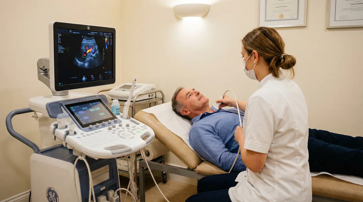
The carotid arteries run up either side of the neck and supply most of the front of the brain. A carotid ultrasound uses Doppler to measure how fast blood is moving through them and to look for fatty plaque narrowing the channel. A tight narrowing is a treatable stroke risk, so this is a standard part of the work-up rather than an optional extra.
Longer heart rhythm monitoring
This is the test that most often changes the answer. Because AF can be intermittent, a 12-lead ECG lasting a few seconds — or even a 24-hour Holter monitor — may record nothing at all while the rhythm is behaving. Monitoring over weeks rather than hours detects AF in a meaningful proportion of people initially labelled cryptogenic; published series put it at up to a quarter.
NICE has assessed implantable cardiac monitors for exactly this situation, recommending the Reveal LINQ device as an option after cryptogenic stroke including TIA, but only where non-invasive ECG monitoring has already been carried out and a rhythm cause is still suspected. That sequencing matters: the simpler tests come first.
From our practice
From our practice
People come to us after a stroke asking a version of the same question: can you find what the hospital missed?
Our honest answer is that we can look carefully at the cardiac side. We can assess the heart for a source such as atrial fibrillation, a valve problem, a clot, or a patent foramen ovale. Those are real, findable things, and identifying one may change what your stroke team recommends.
What we cannot do is prove that a stroke was definitely caused by the heart when the wider stroke work-up is incomplete. A cardiac finding is one piece of a picture that also includes brain imaging, the neck and intracranial arteries, blood tests for clotting disorders, and the pattern of the stroke itself. A PFO found on a scan, for instance, is a common incidental finding in the general population — it does not automatically explain a stroke. Interpreting it needs the whole picture, which is why we report findings back to your stroke or cardiology team rather than presenting them as a standalone answer.
Drawn from general patterns among the patients we see, not any individual case. Every finding still needs individual professional assessment alongside your full stroke work-up.
What these tests cannot tell you
Being clear about the limits is more useful than overselling the tests:
A normal echocardiogram does not exclude a cardiac cause. Intermittent AF leaves no trace on a structural scan.
Finding a PFO is not the same as finding the cause. Around one in four adults has one. Whether it is relevant depends on age, the pattern of the stroke, and whether everything else has been excluded.
Ultrasound does not image the coronary arteries or the small vessels in the brain. Different questions need different tests.
Nothing here replaces the stroke service. Decisions about anticoagulation, PFO closure or carotid surgery are made by specialist teams with the full record in front of them.
NHS and private pathways
NHS stroke service
Private scan alongside it
Who decides what you need
Stroke consultant and multidisciplinary team
You, or your referring clinician
Typical wait for a follow-up echo
Varies by trust; often several weeks
Usually within a few days
Treatment decisions
Made here
Not made here — findings are reported back
Prolonged or implantable monitoring
Yes, where criteria are met
Ambulatory monitoring only
Cost
Free at the point of use
Echocardiogram £350; carotid ultrasound £235
A private scan is worth considering when you are waiting for an appointment that has been recommended, or when you want a second opinion on a specific structural question. It is not a substitute for the stroke pathway, and we will say so if that is what someone is looking for.
Common questions
How common is a cryptogenic stroke?
Roughly one in four ischaemic strokes is classified as cryptogenic after the initial work-up. The proportion falls as more testing is done, particularly with longer rhythm monitoring.
Will an echocardiogram find the cause of my stroke?
It may. An echocardiogram can identify a clot, a valve problem or impaired heart function, any of which could be relevant. A normal result does not rule out a cardiac cause, because intermittent rhythm problems are not visible on a structural scan.
Should I have my PFO closed?
That decision belongs to a cardiologist and stroke specialist working together. It depends on your age, the nature of the stroke, and whether other causes have been excluded. Finding a PFO on a scan is the beginning of that conversation, not the end of it.
How long should heart rhythm monitoring last?
There is no single agreed answer. Detection improves with longer monitoring, but the right duration and device depend on how likely AF is thought to be, and on what you can tolerate. Your cardiologist will weigh that up with you.
Can I self-refer for these scans?
Yes, you can book an echocardiogram or carotid ultrasound with us directly. We would encourage you to tell your stroke team you are doing so, and we will send the report to them if you ask us to.
When will I get my results?
You receive instant verbal results at the appointment from the consultant performing your scan, with the written report following within 24 hours.
CQC-registered clinic · BMUS member · ICO-registered. Reports can be shared securely with your NHS GP or specialist at your request.
References
National Institute for Health and Care Excellence. Stroke and transient ischaemic attack in over 16s: diagnosis and initial management (NG128). Accessed 30 July 2026.
National Institute for Health and Care Excellence. Implantable cardiac monitors to detect atrial fibrillation after cryptogenic stroke (DG41). Accessed 30 July 2026.
National Institute for Health and Care Excellence. Atrial fibrillation: diagnosis and management (NG196). Accessed 30 July 2026.
Gladstone DJ et al. Atrial fibrillation in patients with cryptogenic stroke. New England Journal of Medicine 2014;370:2467–77.