Echo vs CT: Which Heart Test Answers Your Question?
Echo and CT often get compared because they can both be involved in “heart checks” — but they answer different questions.
An echo (echocardiogram) is a heart ultrasound: it shows movement (pumping) and valves in real time, with no radiation.
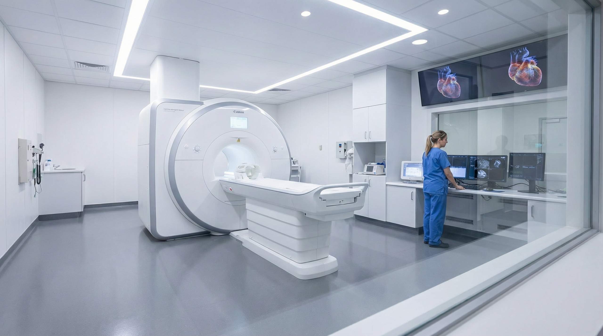
A CT scan uses X-rays to show anatomy in detail (and, in specific pathways, coronary arteries).
This guide helps you choose the right test first, based on symptoms and the decision you’re trying to make.
If you have severe chest pain, collapse, severe breathlessness at rest, or stroke symptoms, seek urgent medical care first.
Private testing is for clarity and next-step guidance — not emergency treatment.
Fast choice: the simplest way to decide
Start with the question you need answered. Don’t start with the technology.
Echo is usually the right first test when your question is:
“How well is my heart pumping?” (function / EF context, wall motion patterns).
“Is a valve causing a murmur or symptoms?” (narrowing/leakage; Doppler flow).
“Could this breathlessness be cardiac?” (in the right clinical context).
“Do I have coronary artery disease?” (in an appropriate pathway such as CT coronary angiography or calcium scoring).
“What does the anatomy look like in detail?” (calcification, specific structural mapping).
“I need an ‘inside view’ that ultrasound can’t provide.” (depending on the clinical question).
CT pathways are typically arranged via GP/cardiology or radiology services. Sonoworld’s cardiovascular imaging service here is echocardiography.
Most common misunderstanding
An echo is not the “blocked arteries test”. A CT pathway may be used for coronary arteries, but it won’t replace the value of echo for valves and real-time function.
Many people need the right one first — and some need both in sequence.
A good echo is structured: chambers, function, valves, Doppler flow, pericardium — so the report answers real decisions (reassurance, referral, monitoring, or a different test).
When CT is usually the better fit
CT is the “detail anatomy” tool, and in certain clinical pathways it can be used to evaluate coronary arteries.
It’s not a substitute for echo when the question is valves or real-time function — it’s a different lens.
Common reasons CT enters the conversation
Coronary artery concern (in the appropriate pathway): CT coronary angiography (CTCA) or calcium scoring may be considered by your clinician.
Detailed structural mapping: anatomy where ultrasound windows are limited or where very detailed detail is required.
Calcification: CT can visualise calcium well (context-dependent), which can support certain risk pathways.
If your main symptom is palpitations
That’s often an ECG/monitoring pathway first. CT usually isn’t the first test for rhythm symptoms.
Patients don’t just choose tests based on “accuracy” — they also care about safety, comfort, and whether the result changes what happens next.
Here’s the straightforward safety picture.
Echo safety profile
No ionising radiation (it’s ultrasound).
Generally very well tolerated. You may feel mild pressure from the probe.
Limits are usually “information limits” (image windows, complexity), not safety limits.
Ionising radiation: CT uses X-rays, so dose is a real consideration (your clinician balances benefit vs risk).
Contrast dye (some pathways): CT coronary angiography may use iodinated contrast; clinicians consider allergy history and kidney function.
Not always the first step: if the question is valves or real-time pumping, echo usually answers more directly.
Always tell your clinician if you’re pregnant, have had a contrast reaction, or have known kidney disease.
The “best test” is the test that changes the next decision
If the result won’t change what you do next, it’s the wrong test (or the wrong time for it).
That’s why symptom → question → test is a better sequence than “test shopping”.
FAQs: echo vs CT
Short answers to the questions people ask right before they book.
Does an echo show blocked coronary arteries?
Not directly. Echo shows structure and function (valves, pumping, chamber size, pericardium). It may show consequences of coronary disease in some situations,
but coronary artery assessment is usually a different pathway (often CT coronary angiography or other clinician-led tests).
Does CT replace an echo?
No. CT is a detailed anatomy tool and may be used for coronary pathways in the right context. Echo is the main test for valves and real-time function.
They can be complementary, but they are not interchangeable.
I’m breathless on exertion — echo or CT?
Echo is often the more direct first step when the question is pumping function, valves, or cardiac contribution to symptoms.
CT may be considered later if the clinical question is coronary arteries or specific detailed anatomy.
If I have palpitations, should I do CT?
Palpitations are usually an ECG/monitoring pathway first (rhythm capture). Echo may be added to check structure and function.
CT is not typically the first test for rhythm symptoms.
Is CT “dangerous” because of radiation?
CT uses ionising radiation, so clinicians weigh benefit vs risk. When the clinical benefit is strong, CT can be an appropriate choice.
If the question can be answered safely and effectively with echo, echo is often preferred.