
Private Heart Health Check and Echocardiogram in London
Feeling your heart race unexpectedly, noticing a tightness across your chest, or finding yourself short of breath during activities that never used to trouble you — these experiences can be deeply unsettling. The worry that follows is often made worse by long NHS waiting lists, where weeks or even months can pass before you receive a diagnostic appointment. You deserve answers quickly, and you deserve them from experienced specialists who take the time to listen.
At Sonoworld Diagnostic Services, based in London's renowned Harley Street medical district, we offer private heart health checks and echocardiograms that give you a thorough assessment of your cardiovascular health — often on the same day you contact us. Our approach is guided by recognised clinical standards, including NICE cardiovascular guidelines, and every assessment is carried out by experienced practitioners with over 20 years of clinical expertise. No GP referral is needed. You simply book, attend, and receive clear answers.
Why Heart Health Screening Matters in the UK
Cardiovascular disease (CVD) remains the leading cause of premature death in the United Kingdom. The British Heart Foundation reports that approximately 7.6 million people across the UK are currently living with heart and circulatory diseases[1]. CVD accounts for one in four premature deaths in England, and coronary heart disease alone is responsible for around 63,000 deaths each year[2]. More concerning still, cardiovascular deaths among working-age adults have risen by 18% since 2019[1].
These figures are not abstract. They represent real people — parents, partners, colleagues — many of whom had no idea they were at risk until a serious event occurred. A private heart health check provides an opportunity to identify risk factors and early signs of cardiovascular disease before they progress. Early detection through screening can lead to lifestyle changes, medication, or monitoring that significantly reduces the chance of a heart attack, stroke, or heart failure.
Who Should Consider a Heart Health Check?
Have you ever wondered whether your symptoms are something to worry about, or whether your family history puts you at higher risk? A heart health check is appropriate for a wide range of people, not only those with obvious symptoms.
Individuals who experience chest discomfort, breathlessness on exertion, palpitations, dizziness, or unexplained fatigue should seek a cardiovascular assessment. Those with a family history of heart disease, high blood pressure, elevated cholesterol, or diabetes also benefit from proactive screening. Smokers, people who lead a sedentary lifestyle, and anyone over the age of 40 who has not had a recent cardiovascular review should consider booking an appointment.
The NHS Health Check programme offers a basic assessment for adults aged 40 to 74 every five years. A private heart health check goes further, providing detailed imaging and specialist interpretation that the standard NHS check does not include.
What is an Echocardiogram?
An echocardiogram, often called an "echo" or a cardiac ultrasound-scan, is a painless, non-invasive imaging test that uses high-frequency sound waves to produce detailed, real-time moving pictures of your heart. Unlike X-rays or CT scans, an echocardiogram does not use any ionising radiation, making it one of the safest diagnostic tools available.
The test allows your clinician to observe the heart's chambers, walls, valves, and surrounding structures as they move. Blood flow patterns through the heart and its major vessels are also assessed using a technique called Doppler imaging. The result is a comprehensive picture of how well your heart is functioning — its size, its shape, the thickness of its walls, and the efficiency of its pumping action.
According to the Cleveland Clinic, "An echocardiogram uses high-frequency sound waves to create moving pictures of your heart as it beats. This test can help your healthcare provider diagnose and treat many different heart conditions."[3]

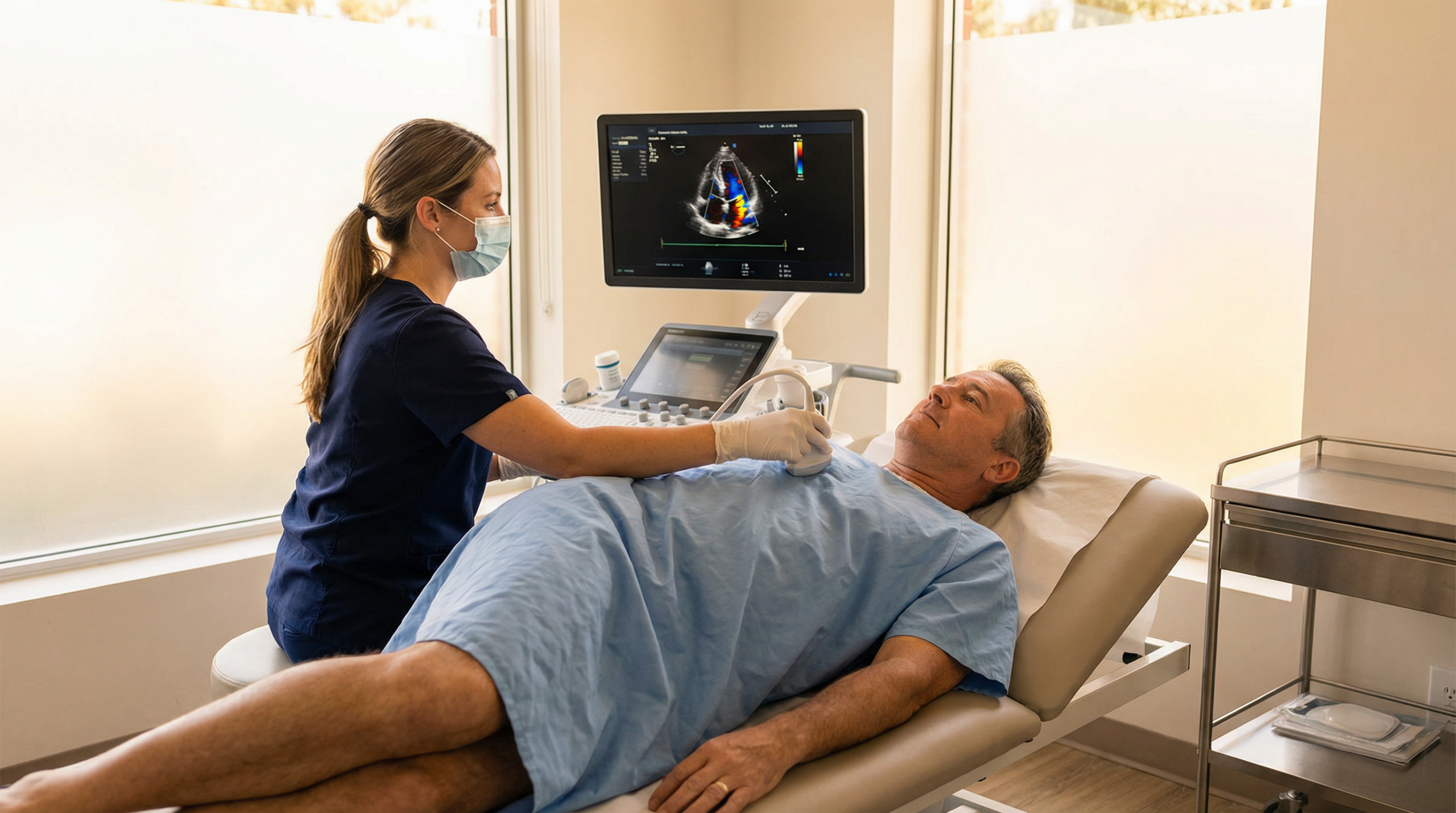
How an Echocardiogram Works
During a standard transthoracic echocardiogram (TTE) — the most common type — you lie on an examination table, usually on your left side. A small amount of gel is applied to your chest, and a handheld device called a transducer is moved across different areas of your chest wall. The transducer emits sound waves that bounce off the structures of your heart, creating echoes. These echoes are captured and converted into moving images on a screen.
The entire process typically takes between 30 and 60 minutes. You remain fully clothed from the waist down, and there are no needles, no injections, and no recovery time. You can return to your normal activities immediately afterwards.
Several imaging techniques may be used during your echocardiogram to gather the most complete information:
| Imaging Technique | What it Shows |
|---|---|
| Two-dimensional (2D) imaging | Cross-sectional "slices" of the heart, showing its structure in real time. |
| Three-dimensional (3D) imaging | A volumetric view of the heart, useful for assessing valve anatomy and chamber size with greater accuracy. |
| Doppler imaging | The speed and direction of blood flow through the heart's chambers and valves. |
| Colour-flow Doppler | A colour-coded map of blood flow, making it easy to identify areas of turbulence or regurgitation. |
| Strain imaging | Changes in how the heart muscle contracts and relaxes, detecting early signs of dysfunction. |
What Can an Echocardiogram Diagnose?
An echocardiogram is one of the most versatile cardiac diagnostic tools. It can identify and help monitor a wide range of heart conditions:
Valve disease is one of the most common findings. The echo can show whether valves are narrowed (stenosis), leaking (regurgitation), or not closing properly (prolapse). Conditions such as mitral valve prolapse, aortic stenosis, and tricuspid regurgitation are all clearly visible on an echocardiogram.
Cardiomyopathy, a disease of the heart muscle, causes the heart to become enlarged, thickened, or stiff. An echo reveals these structural changes and measures how effectively the heart is pumping blood.
Heart failure is assessed by measuring the ejection fraction — the percentage of blood the left ventricle pumps out with each beat. A normal ejection fraction falls between 55% and 70%[4]. Values below 40% indicate significant impairment.
Pericardial disease, including pericarditis (inflammation of the sac around the heart) and pericardial effusion (fluid accumulation), is readily detected. The echo can show the amount of fluid present and whether it is placing pressure on the heart.
Congenital heart defects — structural abnormalities present from birth — such as atrial septal defects (holes between the upper chambers) or ventricular septal defects, are identified through echocardiographic imaging.
Blood clots and cardiac masses, including thrombi within the heart chambers and, rarely, cardiac tumours, can be visualised during the examination.
Infective endocarditis, an infection of the heart valves, produces characteristic growths called vegetations that are visible on echo imaging.
Understanding the Difference: Echocardiogram, ECG, and Holter Monitor
When your doctor mentions heart tests, three names come up most frequently: the echocardiogram, the ECG, and the Holter monitor. Each serves a distinct purpose, and understanding the difference helps you know which test — or combination of tests — is right for your situation. Our guide on echo vs ECG explains this in greater detail.

The ECG (Electrocardiogram)
An ECG records the electrical signals that trigger each heartbeat. Small adhesive electrodes are placed on your chest, arms, and legs, and the test takes just five to ten minutes. The resulting trace — a series of waves and intervals on paper or screen — reveals your heart's rate, rhythm, and electrical conduction pattern.
An ECG is particularly useful for detecting arrhythmias (irregular heart rhythms), signs of a previous heart attack, and abnormalities in the heart's electrical pathways. If you've ever wondered what an abnormal ECG means, it can indicate anything from a benign variation to a condition requiring further investigation.
The limitation of a standard ECG is that it captures only a brief snapshot — typically 10 seconds of your heart's electrical activity. If your symptoms are intermittent, the ECG may appear entirely normal.
The Holter Monitor
A Holter monitor addresses this limitation. It is a portable device that records your heart's electrical activity continuously over 24 to 48 hours (or sometimes longer) while you go about your daily life. The device is small enough to wear under your clothing, and you keep a diary of your activities and any symptoms you experience during the monitoring period.
The Holter monitor is the test of choice when symptoms such as palpitations, dizziness, or fainting episodes occur sporadically. By correlating your symptom diary with the recorded heart rhythm data, your clinician can determine whether your symptoms coincide with an arrhythmia. Our guide on ECG vs Holter monitoring provides a detailed comparison.
Comparing the Three Tests
| Feature | Echocardiogram | ECG | Holter Monitor |
|---|---|---|---|
| What it assesses | Heart structure, muscle function, valve performance, blood flow | Heart's electrical activity, rhythm, and rate | Continuous electrical rhythm recording over 24–48+ hours |
| Technology | Ultrasound (sound waves) | Electrical sensors (electrodes) | Portable continuous ECG recorder |
| Duration | 30–60 minutes | 5–10 minutes | 24–48 hours (or longer) |
| Best for | Structural problems, valve disease, heart failure, cardiomyopathy | Arrhythmias, conduction abnormalities, signs of ischaemia | Intermittent palpitations, unexplained dizziness, sporadic arrhythmias |
| Invasiveness | Non-invasive | Non-invasive | Non-invasive (worn externally) |
| Radiation | None | None | None |
In many cases, your clinician may recommend more than one of these tests. An ECG might be performed first as a quick screening tool, followed by an echocardiogram for a structural assessment, and a Holter monitor if intermittent rhythm disturbances are suspected.
What Happens During a Private Heart Health Check at Sonoworld?
Knowing what to expect before you arrive can ease any apprehension. At Sonoworld, we have designed our heart health check to be thorough, unhurried, and completely patient-focused.
Before Your Appointment
When you book your appointment — by telephone on 020 3633 4902 or through our online booking system — our team will confirm the details and let you know if any specific preparation is needed. For a standard echocardiogram, there is no fasting requirement and no need to stop any medications. You can eat, drink, and take your usual medicines as normal.
Wear comfortable, loose-fitting clothing. You will be asked to undress from the waist up and may be given a gown for the examination.
During Your Appointment
Your appointment begins with a discussion about your symptoms, medical history, and any specific concerns you have. This conversation is not rushed; we take the time to understand your situation fully.
For the echocardiogram itself, you will lie on an examination table, typically on your left side. The practitioner applies a water-based gel to your chest and uses a transducer to capture images of your heart from several different angles. You may be asked to breathe in, breathe out, or hold your breath briefly at certain points to optimise the image quality. The gel may feel slightly cool, but the procedure itself is entirely painless.
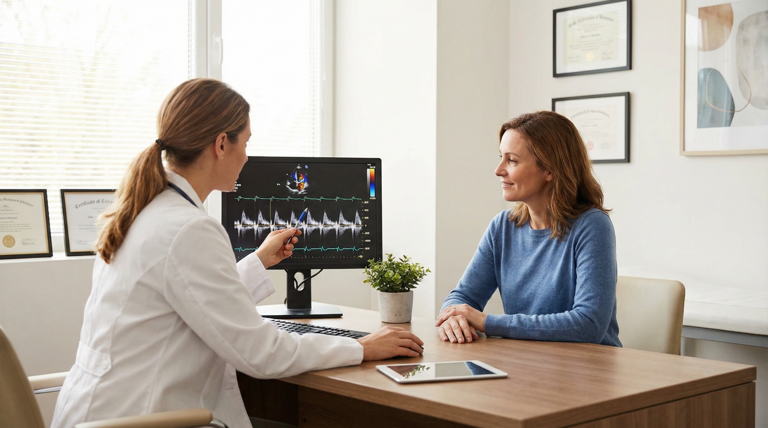
Throughout the examination, your practitioner will explain what they are seeing on the screen. At Sonoworld, we believe in transparency — you will receive immediate verbal feedback during your appointment, so you leave with a clear understanding of your results.
After Your Appointment
Following your echocardiogram, you will receive a comprehensive written report within 24 hours. This report includes detailed measurements, clinical findings, and — where appropriate — recommendations for follow-up. The report can be shared with your GP or referring consultant if you wish.
There is no recovery period. You can drive, return to work, or carry on with your day immediately after the appointment.
The Echocardiogram vs Other Heart Imaging Tests
Patients sometimes ask how an echocardiogram compares with other cardiac imaging methods. Our detailed guide on echo vs CT explores this topic further.
Echocardiogram vs Cardiac CT: A cardiac CT scan uses X-rays and contrast dye to create detailed images of the coronary arteries and heart structures. It is particularly useful for detecting calcium build-up in the arteries (coronary artery calcium scoring) and for visualising the coronary arteries themselves. An echocardiogram, by contrast, excels at assessing the heart's pumping function, valve performance, and blood flow in real time — without radiation or contrast dye.
Echocardiogram vs Cardiac MRI: Cardiac MRI provides exceptionally detailed images of the heart muscle and can characterise tissue (for example, identifying scarring or inflammation). It is typically used for more complex diagnostic questions. An echocardiogram is faster, more widely available, and serves as the first-line imaging test for most cardiac assessments.
Echocardiogram vs ECG: While an ECG records electrical activity, an echocardiogram provides a visual, structural assessment. The two tests are complementary rather than interchangeable.
Conditions and Symptoms That Require an Echocardiogram
Our guide on symptoms that require an echo provides an in-depth look at this topic. The following are among the most common reasons a private echocardiogram is requested:
Chest pain or tightness — whether it occurs at rest, during physical activity, or in response to stress — warrants investigation. While chest pain has many possible causes, an echocardiogram can rule out or confirm cardiac involvement.
Breathlessness that is new, worsening, or disproportionate to the level of exertion may indicate heart failure, valve disease, or pericardial effusion. The echo provides direct evidence of whether the heart is the source.
Heart murmurs detected during a physical examination are often investigated with an echocardiogram. A murmur is an extra sound caused by turbulent blood flow, and the echo can determine whether it is caused by a valve abnormality.
Palpitations and arrhythmias — while primarily assessed with an ECG or Holter monitor — may also prompt an echocardiogram to check for underlying structural causes.
High blood pressure over a prolonged period can cause the heart muscle to thicken (left ventricular hypertrophy). An echocardiogram measures wall thickness and detects this change.
A family history of cardiomyopathy or sudden cardiac death is a strong indication for screening echocardiography, even in the absence of symptoms.
Pre-operative assessment — some patients require an echocardiogram before surgery to confirm that the heart can safely tolerate anaesthesia and the physiological stress of an operation.
Safety, Limitations, and What to Know
An echocardiogram is one of the safest medical imaging tests available. There is no radiation exposure, no contrast dye (in a standard TTE), and no known side effects. The test can be performed on patients of all ages, including pregnant women and children. Our page on echo safety and risks covers this topic in full.
Like all diagnostic tools, the echocardiogram has certain limitations. Image quality can be affected by body habitus — patients with a larger chest wall or significant lung disease may produce images that are more difficult to interpret. In these cases, alternative imaging such as a transesophageal echocardiogram (TEE) or cardiac MRI may be recommended.
An echocardiogram does not directly visualise the coronary arteries. If coronary artery disease is suspected, additional tests such as a cardiac CT, coronary angiography, or a stress test may be needed.
Why Choose Sonoworld for Your Heart Health Check?

Sonoworld Diagnostic Services is a CQC-registered clinic rated "Good," located at 29 Weymouth Street in London's Marylebone — within the prestigious Harley Street medical district. Our lead clinician, Daniela Stan MSc, is a Consultant Ultrasound Practitioner with over two decades of experience across NHS and private practice.
Your Heart Health: A Broader Perspective
A heart health check is often the starting point for a wider cardiovascular assessment. Depending on your risk profile and findings, your clinician may recommend additional investigations:
A carotid artery ultrasound-scan assesses the blood vessels in your neck that supply the brain, screening for narrowing (stenosis) that increases stroke risk.
A DVT ultrasound-scan checks for blood clots in the deep veins of the legs — a condition that, if untreated, can lead to a pulmonary embolism.
An abdominal aortic aneurysm (AAA) ultrasound-scan screens for a dangerous enlargement of the main artery in the abdomen, which often produces no symptoms until it becomes a medical emergency.
Our Well Man and Well Woman screening packages combine multiple assessments into a single, comprehensive health review.
Frequently Asked Questions
A standard transthoracic echocardiogram at Sonoworld typically takes between 30 and 60 minutes, depending on the complexity of the assessment.
No. The test is completely painless. You may feel slight pressure from the transducer and a cool sensation from the gel, but there is no discomfort.
Yes. For a standard transthoracic echocardiogram, there are no dietary restrictions. Eat and drink as you normally would.
At Sonoworld, you receive immediate verbal feedback during your appointment. A full written report is delivered within 24 hours.
A normal left ventricular ejection fraction is between 55% and 70%. Values between 41% and 54% indicate mild to moderate reduction, and values below 40% suggest significant impairment.
No. You can self-refer directly to Sonoworld without a GP referral. Simply book your appointment by phone or online.
Yes. Sonoworld accepts private medical insurance from several providers, including AXA, Healix, and WPA.
Related Topics
Book Your Private Heart Health Check
If you have been experiencing symptoms that concern you, or if you simply want the reassurance that comes with a thorough cardiovascular assessment, our team is ready to help. Contact Sonoworld today to arrange your private heart health check or echocardiogram at our London clinic.
Call us on 020 3633 4902
Book Your Appointment OnlineSonoworld Diagnostic Services • 29 Weymouth Street, Marylebone, London W1G 7DB
References
- British Heart Foundation. Heart & Circulatory Disease Statistics 2025. bhf.org.uk
- NHS England. Cardiovascular Disease (CVD). england.nhs.uk
- Cleveland Clinic. Echocardiogram: Types & What To Expect. clevelandclinic.org
- American Heart Association. Ejection Fraction Heart Failure Measurement. heart.org


